{"title":"煅烧温度对溶胶-凝胶法制备纳米钴铁氧体结构、形貌、磁性、电学和光学性能的影响","authors":"Md. Farid Ahmed, Afia Yasmin, Bristy Biswas, Md. Lutfor Rahman, Juliya Khanam, Rabeya Jahan Rakhi, Mahmuda Hakim, Md. Sahadat Hossain, Firoz Ahmed, Israt Jahan Lithi and Nahid Sharmin","doi":"10.1039/D5MA00209E","DOIUrl":null,"url":null,"abstract":"<p >Synthesis of cobalt ferrite (CoFe<small><sub>2</sub></small>O<small><sub>4</sub></small>) nanoparticles (NPs) through a sol–gel process is an efficient and cost-effective approach. This process is carried out at different calcination temperatures (500 °C, 600 °C, 700 °C, 800 °C, 900 °C and 1000 °C) using cobalt nitrate [Co(NO<small><sub>3</sub></small>)<small><sub>2</sub></small>·6H<small><sub>2</sub></small>O], ferric nitrate [Fe(NO<small><sub>3</sub></small>)<small><sub>3</sub></small>·9H<small><sub>2</sub></small>O], citric acid (C<small><sub>6</sub></small>H<small><sub>8</sub></small>O<small><sub>7</sub></small>·H<small><sub>2</sub></small>O), glycerol (C<small><sub>3</sub></small>H<small><sub>8</sub></small>O<small><sub>3</sub></small>), and ammonium hydroxide (NH<small><sub>4</sub></small>OH). It is found that different calcination temperatures affect the size of the crystallite produced, <em>i.e.</em>, at higher temperature the size of the crystals increases. To study the structural, optical, magnetic and dielectric properties of the cobalt ferrite nanoparticles synthesized by a sol–gel method, characterization techniques such as X-ray diffraction (XRD), simultaneous thermal analysis (STA), vibrating sample magnetometry (VSM), scanning electron microscopy (SEM) and Fourier transform infrared (FTIR) spectroscopy were carried out. XRD proved the face centered cubic structure of the NPs. At 1000 °C the sample T6 exhibited a higher zeta potential proving the stability of the solution. The crystallite size and lattice strain were measured by the Debye–Scherrer (D–S) method, Williamson–Hall (W–H) process, Halder–Wagner (H–W) method and size–strain plot (SSP) technique. XRD data confirm the presence of the single spinel phase of cobalt ferrite NPs for all the samples. The sample T1 showed the lowest crystallite size, and the crystallite size ranged from 33 nm to 169 nm for the T6 sample. Two FTIR absorption bands observed at about 402–403 cm<small><sup>−1</sup></small> and 576–580 cm<small><sup>−1</sup></small> are due to the octahedral M–O bond and the M–O bond at the tetrahedral site in the spinel cobalt ferrite, respectively. The SEM micrographs showed that the produced NPs are spherical in shape and homogenously distributed. The average particle size is found to be 46.72 nm for the sample annealed at 800 °C. The maximum saturation magnetization was found to be around 85–62 emu g<small><sup>−1</sup></small>. The band gap energy was found using the Kubelka–Munk method, and it was found that as the annealing temperature increases the size of crystals increases and band gap energy ranges from 3.00 to 3.52 eV, respectively. Thus the sol–gel method can be used to modify the crystallite size at different calcination temperatures.</p>","PeriodicalId":18242,"journal":{"name":"Materials Advances","volume":" 19","pages":" 6724-6741"},"PeriodicalIF":4.7000,"publicationDate":"2025-08-12","publicationTypes":"Journal Article","fieldsOfStudy":null,"isOpenAccess":false,"openAccessPdf":"https://pubs.rsc.org/en/content/articlepdf/2025/ma/d5ma00209e?page=search","citationCount":"0","resultStr":"{\"title\":\"Effect of calcination temperature on nano-cobalt ferrite synthesized by a sol–gel method for modification of its structural, morphological, magnetic, electrical and optical properties\",\"authors\":\"Md. Farid Ahmed, Afia Yasmin, Bristy Biswas, Md. Lutfor Rahman, Juliya Khanam, Rabeya Jahan Rakhi, Mahmuda Hakim, Md. Sahadat Hossain, Firoz Ahmed, Israt Jahan Lithi and Nahid Sharmin\",\"doi\":\"10.1039/D5MA00209E\",\"DOIUrl\":null,\"url\":null,\"abstract\":\"<p >Synthesis of cobalt ferrite (CoFe<small><sub>2</sub></small>O<small><sub>4</sub></small>) nanoparticles (NPs) through a sol–gel process is an efficient and cost-effective approach. This process is carried out at different calcination temperatures (500 °C, 600 °C, 700 °C, 800 °C, 900 °C and 1000 °C) using cobalt nitrate [Co(NO<small><sub>3</sub></small>)<small><sub>2</sub></small>·6H<small><sub>2</sub></small>O], ferric nitrate [Fe(NO<small><sub>3</sub></small>)<small><sub>3</sub></small>·9H<small><sub>2</sub></small>O], citric acid (C<small><sub>6</sub></small>H<small><sub>8</sub></small>O<small><sub>7</sub></small>·H<small><sub>2</sub></small>O), glycerol (C<small><sub>3</sub></small>H<small><sub>8</sub></small>O<small><sub>3</sub></small>), and ammonium hydroxide (NH<small><sub>4</sub></small>OH). It is found that different calcination temperatures affect the size of the crystallite produced, <em>i.e.</em>, at higher temperature the size of the crystals increases. To study the structural, optical, magnetic and dielectric properties of the cobalt ferrite nanoparticles synthesized by a sol–gel method, characterization techniques such as X-ray diffraction (XRD), simultaneous thermal analysis (STA), vibrating sample magnetometry (VSM), scanning electron microscopy (SEM) and Fourier transform infrared (FTIR) spectroscopy were carried out. XRD proved the face centered cubic structure of the NPs. At 1000 °C the sample T6 exhibited a higher zeta potential proving the stability of the solution. The crystallite size and lattice strain were measured by the Debye–Scherrer (D–S) method, Williamson–Hall (W–H) process, Halder–Wagner (H–W) method and size–strain plot (SSP) technique. XRD data confirm the presence of the single spinel phase of cobalt ferrite NPs for all the samples. The sample T1 showed the lowest crystallite size, and the crystallite size ranged from 33 nm to 169 nm for the T6 sample. Two FTIR absorption bands observed at about 402–403 cm<small><sup>−1</sup></small> and 576–580 cm<small><sup>−1</sup></small> are due to the octahedral M–O bond and the M–O bond at the tetrahedral site in the spinel cobalt ferrite, respectively. The SEM micrographs showed that the produced NPs are spherical in shape and homogenously distributed. The average particle size is found to be 46.72 nm for the sample annealed at 800 °C. The maximum saturation magnetization was found to be around 85–62 emu g<small><sup>−1</sup></small>. The band gap energy was found using the Kubelka–Munk method, and it was found that as the annealing temperature increases the size of crystals increases and band gap energy ranges from 3.00 to 3.52 eV, respectively. Thus the sol–gel method can be used to modify the crystallite size at different calcination temperatures.</p>\",\"PeriodicalId\":18242,\"journal\":{\"name\":\"Materials Advances\",\"volume\":\" 19\",\"pages\":\" 6724-6741\"},\"PeriodicalIF\":4.7000,\"publicationDate\":\"2025-08-12\",\"publicationTypes\":\"Journal Article\",\"fieldsOfStudy\":null,\"isOpenAccess\":false,\"openAccessPdf\":\"https://pubs.rsc.org/en/content/articlepdf/2025/ma/d5ma00209e?page=search\",\"citationCount\":\"0\",\"resultStr\":null,\"platform\":\"Semanticscholar\",\"paperid\":null,\"PeriodicalName\":\"Materials Advances\",\"FirstCategoryId\":\"1085\",\"ListUrlMain\":\"https://pubs.rsc.org/en/content/articlelanding/2025/ma/d5ma00209e\",\"RegionNum\":0,\"RegionCategory\":null,\"ArticlePicture\":[],\"TitleCN\":null,\"AbstractTextCN\":null,\"PMCID\":null,\"EPubDate\":\"\",\"PubModel\":\"\",\"JCR\":\"Q2\",\"JCRName\":\"MATERIALS SCIENCE, MULTIDISCIPLINARY\",\"Score\":null,\"Total\":0}","platform":"Semanticscholar","paperid":null,"PeriodicalName":"Materials Advances","FirstCategoryId":"1085","ListUrlMain":"https://pubs.rsc.org/en/content/articlelanding/2025/ma/d5ma00209e","RegionNum":0,"RegionCategory":null,"ArticlePicture":[],"TitleCN":null,"AbstractTextCN":null,"PMCID":null,"EPubDate":"","PubModel":"","JCR":"Q2","JCRName":"MATERIALS SCIENCE, MULTIDISCIPLINARY","Score":null,"Total":0}
Effect of calcination temperature on nano-cobalt ferrite synthesized by a sol–gel method for modification of its structural, morphological, magnetic, electrical and optical properties
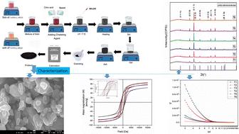
Synthesis of cobalt ferrite (CoFe2O4) nanoparticles (NPs) through a sol–gel process is an efficient and cost-effective approach. This process is carried out at different calcination temperatures (500 °C, 600 °C, 700 °C, 800 °C, 900 °C and 1000 °C) using cobalt nitrate [Co(NO3)2·6H2O], ferric nitrate [Fe(NO3)3·9H2O], citric acid (C6H8O7·H2O), glycerol (C3H8O3), and ammonium hydroxide (NH4OH). It is found that different calcination temperatures affect the size of the crystallite produced, i.e., at higher temperature the size of the crystals increases. To study the structural, optical, magnetic and dielectric properties of the cobalt ferrite nanoparticles synthesized by a sol–gel method, characterization techniques such as X-ray diffraction (XRD), simultaneous thermal analysis (STA), vibrating sample magnetometry (VSM), scanning electron microscopy (SEM) and Fourier transform infrared (FTIR) spectroscopy were carried out. XRD proved the face centered cubic structure of the NPs. At 1000 °C the sample T6 exhibited a higher zeta potential proving the stability of the solution. The crystallite size and lattice strain were measured by the Debye–Scherrer (D–S) method, Williamson–Hall (W–H) process, Halder–Wagner (H–W) method and size–strain plot (SSP) technique. XRD data confirm the presence of the single spinel phase of cobalt ferrite NPs for all the samples. The sample T1 showed the lowest crystallite size, and the crystallite size ranged from 33 nm to 169 nm for the T6 sample. Two FTIR absorption bands observed at about 402–403 cm−1 and 576–580 cm−1 are due to the octahedral M–O bond and the M–O bond at the tetrahedral site in the spinel cobalt ferrite, respectively. The SEM micrographs showed that the produced NPs are spherical in shape and homogenously distributed. The average particle size is found to be 46.72 nm for the sample annealed at 800 °C. The maximum saturation magnetization was found to be around 85–62 emu g−1. The band gap energy was found using the Kubelka–Munk method, and it was found that as the annealing temperature increases the size of crystals increases and band gap energy ranges from 3.00 to 3.52 eV, respectively. Thus the sol–gel method can be used to modify the crystallite size at different calcination temperatures.

 求助内容:
求助内容: 应助结果提醒方式:


