黄斑毛细血管扩张伴内限制膜缺损的2型黄斑孔的外科治疗及勒细胞功能的影像学评价。

IF 2.6 3区 医学 Q2 ONCOLOGY
Nianjia Wang , Jiayi Wu , Xintong Xiang , Ke Wu , Yujia Gao , Liang Yao , Yuping Zheng
{"title":"黄斑毛细血管扩张伴内限制膜缺损的2型黄斑孔的外科治疗及<s:1>勒细胞功能的影像学评价。","authors":"Nianjia Wang ,&nbsp;Jiayi Wu ,&nbsp;Xintong Xiang ,&nbsp;Ke Wu ,&nbsp;Yujia Gao ,&nbsp;Liang Yao ,&nbsp;Yuping Zheng","doi":"10.1016/j.pdpdt.2025.105236","DOIUrl":null,"url":null,"abstract":"<div><div>This study presents a rare case of macular telangiectasia type 2 (Mac Tel 2) with a secondary full-thickness macular hole, in which an unexpected internal limiting membrane (ILM) deficiency was discovered intraoperatively. Swept-source OCT angiography (SS-OCTA) volumetric data were utilized to construct 3D models for the assessment of Müller cell function. Although ILM flap surgery achieved initial anatomical closure and visual improvement, the hole reopened after six years, with corresponding vision decline. Intraoperative ICG staining revealed patchy ILM absence, indicating severe Müller cell dysfunction. 3D vascular and structural analysis demonstrated topographic collapse correlating with loss of cytoskeletal support from Müller cells, explaining the temporary surgical success and subsequent recurrence. The findings suggest that long-term closure of Mac Tel 2-associated macular holes depends on underlying disease progression rather than surgical technique alone. For eyes with advanced Müller cell degeneration, alternative closure materials should be considered. This case highlights the critical role of vascular and structural 3D imaging in evaluating cellular function and predicting surgical outcomes.</div></div>","PeriodicalId":20141,"journal":{"name":"Photodiagnosis and Photodynamic Therapy","volume":"56 ","pages":"Article 105236"},"PeriodicalIF":2.6000,"publicationDate":"2025-09-25","publicationTypes":"Journal Article","fieldsOfStudy":null,"isOpenAccess":false,"openAccessPdf":"","citationCount":"0","resultStr":"{\"title\":\"Surgical management of macular telangiectasia type 2-associated macular hole with internal limiting membrane deficiency and imaging assessment of müller cell function\",\"authors\":\"Nianjia Wang ,&nbsp;Jiayi Wu ,&nbsp;Xintong Xiang ,&nbsp;Ke Wu ,&nbsp;Yujia Gao ,&nbsp;Liang Yao ,&nbsp;Yuping Zheng\",\"doi\":\"10.1016/j.pdpdt.2025.105236\",\"DOIUrl\":null,\"url\":null,\"abstract\":\"<div><div>This study presents a rare case of macular telangiectasia type 2 (Mac Tel 2) with a secondary full-thickness macular hole, in which an unexpected internal limiting membrane (ILM) deficiency was discovered intraoperatively. Swept-source OCT angiography (SS-OCTA) volumetric data were utilized to construct 3D models for the assessment of Müller cell function. Although ILM flap surgery achieved initial anatomical closure and visual improvement, the hole reopened after six years, with corresponding vision decline. Intraoperative ICG staining revealed patchy ILM absence, indicating severe Müller cell dysfunction. 3D vascular and structural analysis demonstrated topographic collapse correlating with loss of cytoskeletal support from Müller cells, explaining the temporary surgical success and subsequent recurrence. The findings suggest that long-term closure of Mac Tel 2-associated macular holes depends on underlying disease progression rather than surgical technique alone. For eyes with advanced Müller cell degeneration, alternative closure materials should be considered. This case highlights the critical role of vascular and structural 3D imaging in evaluating cellular function and predicting surgical outcomes.</div></div>\",\"PeriodicalId\":20141,\"journal\":{\"name\":\"Photodiagnosis and Photodynamic Therapy\",\"volume\":\"56 \",\"pages\":\"Article 105236\"},\"PeriodicalIF\":2.6000,\"publicationDate\":\"2025-09-25\",\"publicationTypes\":\"Journal Article\",\"fieldsOfStudy\":null,\"isOpenAccess\":false,\"openAccessPdf\":\"\",\"citationCount\":\"0\",\"resultStr\":null,\"platform\":\"Semanticscholar\",\"paperid\":null,\"PeriodicalName\":\"Photodiagnosis and Photodynamic Therapy\",\"FirstCategoryId\":\"3\",\"ListUrlMain\":\"https://www.sciencedirect.com/science/article/pii/S1572100025007677\",\"RegionNum\":3,\"RegionCategory\":\"医学\",\"ArticlePicture\":[],\"TitleCN\":null,\"AbstractTextCN\":null,\"PMCID\":null,\"EPubDate\":\"\",\"PubModel\":\"\",\"JCR\":\"Q2\",\"JCRName\":\"ONCOLOGY\",\"Score\":null,\"Total\":0}","platform":"Semanticscholar","paperid":null,"PeriodicalName":"Photodiagnosis and Photodynamic Therapy","FirstCategoryId":"3","ListUrlMain":"https://www.sciencedirect.com/science/article/pii/S1572100025007677","RegionNum":3,"RegionCategory":"医学","ArticlePicture":[],"TitleCN":null,"AbstractTextCN":null,"PMCID":null,"EPubDate":"","PubModel":"","JCR":"Q2","JCRName":"ONCOLOGY","Score":null,"Total":0}
引用次数: 0

摘要

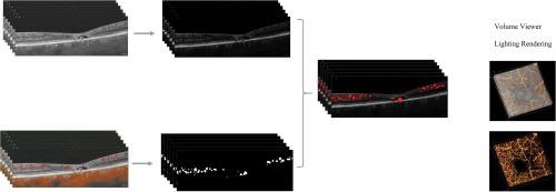
本研究报告一例罕见的黄斑毛细血管扩张2型(Mac Tel 2)伴继发性全层黄斑孔,术中发现意外的内限制膜(ILM)缺陷。扫描源OCT血管造影(SS-OCTA)体积数据用于构建三维模型以评估心肌细胞功能。虽然ILM皮瓣手术获得了初步的解剖闭合和视力改善,但6年后空洞重新开放,视力相应下降。术中ICG染色显示斑片状ILM缺失,提示严重的颞叶细胞功能障碍。三维血管和结构分析表明,地形塌陷与勒细胞骨架支持的丧失有关,解释了暂时手术成功和随后的复发。研究结果表明,Mac - Tel - 2相关黄斑孔的长期闭合取决于潜在的疾病进展,而不仅仅是手术技术。对于晚期勒细胞变性的眼睛,应考虑其他闭合材料。本病例强调了血管和结构三维成像在评估细胞功能和预测手术结果中的关键作用。
本文章由计算机程序翻译,如有差异,请以英文原文为准。

Surgical management of macular telangiectasia type 2-associated macular hole with internal limiting membrane deficiency and imaging assessment of müller cell function

Surgical management of macular telangiectasia type 2-associated macular hole with internal limiting membrane deficiency and imaging assessment of müller cell function
This study presents a rare case of macular telangiectasia type 2 (Mac Tel 2) with a secondary full-thickness macular hole, in which an unexpected internal limiting membrane (ILM) deficiency was discovered intraoperatively. Swept-source OCT angiography (SS-OCTA) volumetric data were utilized to construct 3D models for the assessment of Müller cell function. Although ILM flap surgery achieved initial anatomical closure and visual improvement, the hole reopened after six years, with corresponding vision decline. Intraoperative ICG staining revealed patchy ILM absence, indicating severe Müller cell dysfunction. 3D vascular and structural analysis demonstrated topographic collapse correlating with loss of cytoskeletal support from Müller cells, explaining the temporary surgical success and subsequent recurrence. The findings suggest that long-term closure of Mac Tel 2-associated macular holes depends on underlying disease progression rather than surgical technique alone. For eyes with advanced Müller cell degeneration, alternative closure materials should be considered. This case highlights the critical role of vascular and structural 3D imaging in evaluating cellular function and predicting surgical outcomes.
求助全文
通过发布文献求助,成功后即可免费获取论文全文。 去求助
来源期刊
CiteScore
5.80
自引率
24.20%
发文量
509
审稿时长
50 days
期刊介绍: Photodiagnosis and Photodynamic Therapy is an international journal for the dissemination of scientific knowledge and clinical developments of Photodiagnosis and Photodynamic Therapy in all medical specialties. The journal publishes original articles, review articles, case presentations, "how-to-do-it" articles, Letters to the Editor, short communications and relevant images with short descriptions. All submitted material is subject to a strict peer-review process.
×
引用
GB/T 7714-2015
复制
MLA
复制
APA
复制
导出至
BibTeX EndNote RefMan NoteFirst NoteExpress
×
提示
您的信息不完整,为了账户安全,请先补充。
现在去补充
×
提示
您因"违规操作"
具体请查看互助需知
我知道了
×
提示
确定
请完成安全验证×
copy
已复制链接
快去分享给好友吧!
我知道了
右上角分享
点击右上角分享
0
联系我们:info@booksci.cn Book学术提供免费学术资源搜索服务,方便国内外学者检索中英文文献。致力于提供最便捷和优质的服务体验。 Copyright © 2023 布克学术 All rights reserved.
京ICP备2023020795号-1
ghs 京公网安备 11010802042870号
Book学术文献互助
Book学术文献互助群
群 号:604180095
Book学术官方微信