{"title":"基于深度学习的寨卡病毒和SARS-CoV-2线粒体损伤自动分割与分析","authors":"Brianda Alexia Agundis-Tinajero, Miguel Ángel Coronado-Ipiña, Ignacio Lara-Hernández, Rodrigo Aparicio-Antonio, Anita Aguirre-Barbosa, Gisela Barrera-Badillo, Nidia Aréchiga-Ceballos, Irma López-Martínez, Claudia G Castillo, Vanessa Labrada-Martagón, Mauricio Comas-García, Aldo Rodrigo Mejía-Rodríguez","doi":"10.3390/v17091272","DOIUrl":null,"url":null,"abstract":"<p><p>Viruses can induce various mitochondrial morphological changes, which are associated with the type of immune response. Therefore, characterization and analysis of mitochondrial ultrastructural changes could provide insights into the kind of immune response elicited, especially when compared to uninfected cells. However, this analysis is highly time-consuming and susceptible to observer bias. This work presents the development of a deep learning-based approach for the automatic identification, segmentation, and analysis of mitochondria from thin-section transmission electron microscopy images of cells infected with two SARS-CoV-2 variants or the Zika virus, utilizing a convolutional neural network with a U-Net architecture. A comparison between manual and automatic segmentations, along with morphological metrics, was performed, yielding an accuracy greater than 85% with no statistically significant differences between the manual and automatic metrics. This approach significantly reduces processing time and enables a prediction of the immune response to viral infections by allowing the detection of both intact and damaged mitochondria. Therefore, the proposed deep learning-based tool may represent a significant advancement in the study and understanding of cellular responses to emerging pathogens. Additionally, its applicability could be extended to the analysis of other organelles, thereby opening up new opportunities for automated studies in cell biology.</p>","PeriodicalId":49328,"journal":{"name":"Viruses-Basel","volume":"17 9","pages":""},"PeriodicalIF":3.5000,"publicationDate":"2025-09-19","publicationTypes":"Journal Article","fieldsOfStudy":null,"isOpenAccess":false,"openAccessPdf":"https://www.ncbi.nlm.nih.gov/pmc/articles/PMC12474462/pdf/","citationCount":"0","resultStr":"{\"title\":\"Deep Learning-Based Automatic Segmentation and Analysis of Mitochondrial Damage by Zika Virus and SARS-CoV-2.\",\"authors\":\"Brianda Alexia Agundis-Tinajero, Miguel Ángel Coronado-Ipiña, Ignacio Lara-Hernández, Rodrigo Aparicio-Antonio, Anita Aguirre-Barbosa, Gisela Barrera-Badillo, Nidia Aréchiga-Ceballos, Irma López-Martínez, Claudia G Castillo, Vanessa Labrada-Martagón, Mauricio Comas-García, Aldo Rodrigo Mejía-Rodríguez\",\"doi\":\"10.3390/v17091272\",\"DOIUrl\":null,\"url\":null,\"abstract\":\"<p><p>Viruses can induce various mitochondrial morphological changes, which are associated with the type of immune response. Therefore, characterization and analysis of mitochondrial ultrastructural changes could provide insights into the kind of immune response elicited, especially when compared to uninfected cells. However, this analysis is highly time-consuming and susceptible to observer bias. This work presents the development of a deep learning-based approach for the automatic identification, segmentation, and analysis of mitochondria from thin-section transmission electron microscopy images of cells infected with two SARS-CoV-2 variants or the Zika virus, utilizing a convolutional neural network with a U-Net architecture. A comparison between manual and automatic segmentations, along with morphological metrics, was performed, yielding an accuracy greater than 85% with no statistically significant differences between the manual and automatic metrics. This approach significantly reduces processing time and enables a prediction of the immune response to viral infections by allowing the detection of both intact and damaged mitochondria. Therefore, the proposed deep learning-based tool may represent a significant advancement in the study and understanding of cellular responses to emerging pathogens. Additionally, its applicability could be extended to the analysis of other organelles, thereby opening up new opportunities for automated studies in cell biology.</p>\",\"PeriodicalId\":49328,\"journal\":{\"name\":\"Viruses-Basel\",\"volume\":\"17 9\",\"pages\":\"\"},\"PeriodicalIF\":3.5000,\"publicationDate\":\"2025-09-19\",\"publicationTypes\":\"Journal Article\",\"fieldsOfStudy\":null,\"isOpenAccess\":false,\"openAccessPdf\":\"https://www.ncbi.nlm.nih.gov/pmc/articles/PMC12474462/pdf/\",\"citationCount\":\"0\",\"resultStr\":null,\"platform\":\"Semanticscholar\",\"paperid\":null,\"PeriodicalName\":\"Viruses-Basel\",\"FirstCategoryId\":\"3\",\"ListUrlMain\":\"https://doi.org/10.3390/v17091272\",\"RegionNum\":3,\"RegionCategory\":\"医学\",\"ArticlePicture\":[],\"TitleCN\":null,\"AbstractTextCN\":null,\"PMCID\":null,\"EPubDate\":\"\",\"PubModel\":\"\",\"JCR\":\"Q2\",\"JCRName\":\"VIROLOGY\",\"Score\":null,\"Total\":0}","platform":"Semanticscholar","paperid":null,"PeriodicalName":"Viruses-Basel","FirstCategoryId":"3","ListUrlMain":"https://doi.org/10.3390/v17091272","RegionNum":3,"RegionCategory":"医学","ArticlePicture":[],"TitleCN":null,"AbstractTextCN":null,"PMCID":null,"EPubDate":"","PubModel":"","JCR":"Q2","JCRName":"VIROLOGY","Score":null,"Total":0}
Deep Learning-Based Automatic Segmentation and Analysis of Mitochondrial Damage by Zika Virus and SARS-CoV-2.
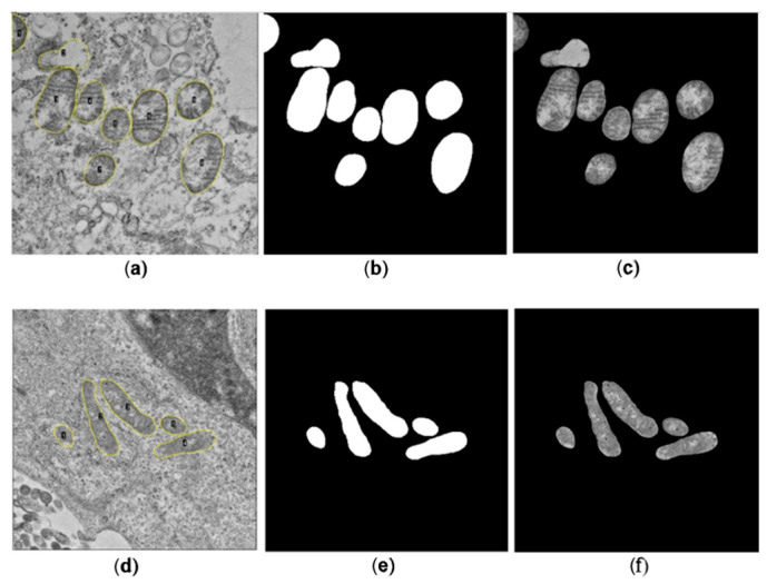
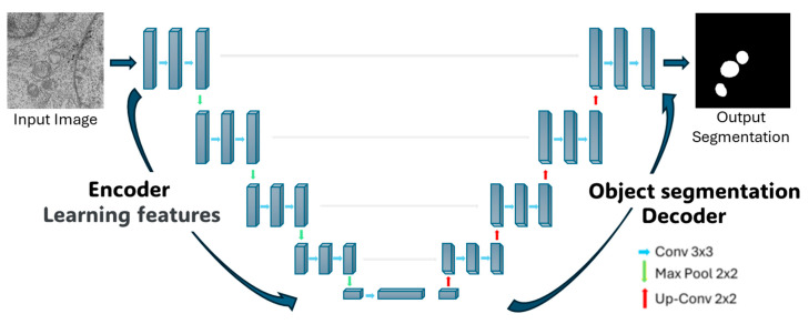
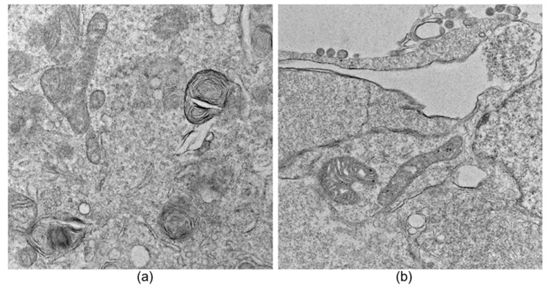
Viruses can induce various mitochondrial morphological changes, which are associated with the type of immune response. Therefore, characterization and analysis of mitochondrial ultrastructural changes could provide insights into the kind of immune response elicited, especially when compared to uninfected cells. However, this analysis is highly time-consuming and susceptible to observer bias. This work presents the development of a deep learning-based approach for the automatic identification, segmentation, and analysis of mitochondria from thin-section transmission electron microscopy images of cells infected with two SARS-CoV-2 variants or the Zika virus, utilizing a convolutional neural network with a U-Net architecture. A comparison between manual and automatic segmentations, along with morphological metrics, was performed, yielding an accuracy greater than 85% with no statistically significant differences between the manual and automatic metrics. This approach significantly reduces processing time and enables a prediction of the immune response to viral infections by allowing the detection of both intact and damaged mitochondria. Therefore, the proposed deep learning-based tool may represent a significant advancement in the study and understanding of cellular responses to emerging pathogens. Additionally, its applicability could be extended to the analysis of other organelles, thereby opening up new opportunities for automated studies in cell biology.
期刊介绍:
Viruses (ISSN 1999-4915) is an open access journal which provides an advanced forum for studies of viruses. It publishes reviews, regular research papers, communications, conference reports and short notes. Our aim is to encourage scientists to publish their experimental and theoretical results in as much detail as possible. There is no restriction on the length of the papers. The full experimental details must be provided so that the results can be reproduced. We also encourage the publication of timely reviews and commentaries on topics of interest to the virology community and feature highlights from the virology literature in the ''News and Views'' section. Electronic files or software regarding the full details of the calculation and experimental procedure, if unable to be published in a normal way, can be deposited as supplementary material.
 求助内容:
求助内容: 应助结果提醒方式:


