Peng Zhou, Jiankang Xuan, Weixiao Xu, Di An, Sining Meng, Hongchao Zhang, Miaoyang Hu, Wanqingyang Hui, Yifei Wang, Jie Cheng, Jianping Xiong, Jun Wang, Xufeng Chen
{"title":"甲基安非他命暴露通过RIPK1-RIPK3-MLKL轴通透线粒体诱导神经元程序性坏死","authors":"Peng Zhou, Jiankang Xuan, Weixiao Xu, Di An, Sining Meng, Hongchao Zhang, Miaoyang Hu, Wanqingyang Hui, Yifei Wang, Jie Cheng, Jianping Xiong, Jun Wang, Xufeng Chen","doi":"10.3390/toxics13090736","DOIUrl":null,"url":null,"abstract":"<p><p>Methamphetamine (Meth), a psychostimulant drug of the amphetamine type, is widely abused and highly neurotoxic. Meth exposure leads to neuronal necroptosis, and the mitochondrial dysfunction may be involved. However, the underlying mechanisms remain poorly understood. Here, we found that Meth significantly elicited the formation of the RIPK1-RIPK3-MLKL necrosome complex. Intriguingly, the activated MLKL (p-MLKL) translocated to the mitochondrial membrane and displayed pore-forming activity, manifesting as the penetration of MLKL in the cell membranes of the mitochondria, which caused decreased mitochondrial membrane potential, ATP generation, and mitochondrial DNA (mtDNA) and increased mitochondrial ROS (mtROS) generation, which finalized neuronal necroptosis. Notably, MLKL activation and translocation seem to depend on the RIPK1-RIPK3 axis since these adverse effects can be substantially ameliorated by disruption of the necrosome complex formation by the necroptotic inhibitor 1 (Nec-1), which also markedly impeded the MLKL mitochondrial membrane translocation. Finally, to delineate the effects of pore formation-associated ROS generation, specific blockage of mtROS retarded the Meth-induced neuronal necroptosis. In conclusion, our study reveals for the first time that MLKL mitochondrial membrane translocation may be involved in Meth-induced neuronal necroptosis. Therefore, impeding MLKL translocation might provide a novel therapeutic strategy for Meth-induced neurotoxicity.</p>","PeriodicalId":23195,"journal":{"name":"Toxics","volume":"13 9","pages":""},"PeriodicalIF":4.1000,"publicationDate":"2025-08-30","publicationTypes":"Journal Article","fieldsOfStudy":null,"isOpenAccess":false,"openAccessPdf":"https://www.ncbi.nlm.nih.gov/pmc/articles/PMC12473512/pdf/","citationCount":"0","resultStr":"{\"title\":\"Methamphetamine Exposure Induces Neuronal Programmed Necrosis by Permeabilizing Mitochondria via the RIPK1-RIPK3-MLKL Axis.\",\"authors\":\"Peng Zhou, Jiankang Xuan, Weixiao Xu, Di An, Sining Meng, Hongchao Zhang, Miaoyang Hu, Wanqingyang Hui, Yifei Wang, Jie Cheng, Jianping Xiong, Jun Wang, Xufeng Chen\",\"doi\":\"10.3390/toxics13090736\",\"DOIUrl\":null,\"url\":null,\"abstract\":\"<p><p>Methamphetamine (Meth), a psychostimulant drug of the amphetamine type, is widely abused and highly neurotoxic. Meth exposure leads to neuronal necroptosis, and the mitochondrial dysfunction may be involved. However, the underlying mechanisms remain poorly understood. Here, we found that Meth significantly elicited the formation of the RIPK1-RIPK3-MLKL necrosome complex. Intriguingly, the activated MLKL (p-MLKL) translocated to the mitochondrial membrane and displayed pore-forming activity, manifesting as the penetration of MLKL in the cell membranes of the mitochondria, which caused decreased mitochondrial membrane potential, ATP generation, and mitochondrial DNA (mtDNA) and increased mitochondrial ROS (mtROS) generation, which finalized neuronal necroptosis. Notably, MLKL activation and translocation seem to depend on the RIPK1-RIPK3 axis since these adverse effects can be substantially ameliorated by disruption of the necrosome complex formation by the necroptotic inhibitor 1 (Nec-1), which also markedly impeded the MLKL mitochondrial membrane translocation. Finally, to delineate the effects of pore formation-associated ROS generation, specific blockage of mtROS retarded the Meth-induced neuronal necroptosis. In conclusion, our study reveals for the first time that MLKL mitochondrial membrane translocation may be involved in Meth-induced neuronal necroptosis. Therefore, impeding MLKL translocation might provide a novel therapeutic strategy for Meth-induced neurotoxicity.</p>\",\"PeriodicalId\":23195,\"journal\":{\"name\":\"Toxics\",\"volume\":\"13 9\",\"pages\":\"\"},\"PeriodicalIF\":4.1000,\"publicationDate\":\"2025-08-30\",\"publicationTypes\":\"Journal Article\",\"fieldsOfStudy\":null,\"isOpenAccess\":false,\"openAccessPdf\":\"https://www.ncbi.nlm.nih.gov/pmc/articles/PMC12473512/pdf/\",\"citationCount\":\"0\",\"resultStr\":null,\"platform\":\"Semanticscholar\",\"paperid\":null,\"PeriodicalName\":\"Toxics\",\"FirstCategoryId\":\"93\",\"ListUrlMain\":\"https://doi.org/10.3390/toxics13090736\",\"RegionNum\":3,\"RegionCategory\":\"环境科学与生态学\",\"ArticlePicture\":[],\"TitleCN\":null,\"AbstractTextCN\":null,\"PMCID\":null,\"EPubDate\":\"\",\"PubModel\":\"\",\"JCR\":\"Q2\",\"JCRName\":\"ENVIRONMENTAL SCIENCES\",\"Score\":null,\"Total\":0}","platform":"Semanticscholar","paperid":null,"PeriodicalName":"Toxics","FirstCategoryId":"93","ListUrlMain":"https://doi.org/10.3390/toxics13090736","RegionNum":3,"RegionCategory":"环境科学与生态学","ArticlePicture":[],"TitleCN":null,"AbstractTextCN":null,"PMCID":null,"EPubDate":"","PubModel":"","JCR":"Q2","JCRName":"ENVIRONMENTAL SCIENCES","Score":null,"Total":0}
Methamphetamine Exposure Induces Neuronal Programmed Necrosis by Permeabilizing Mitochondria via the RIPK1-RIPK3-MLKL Axis.
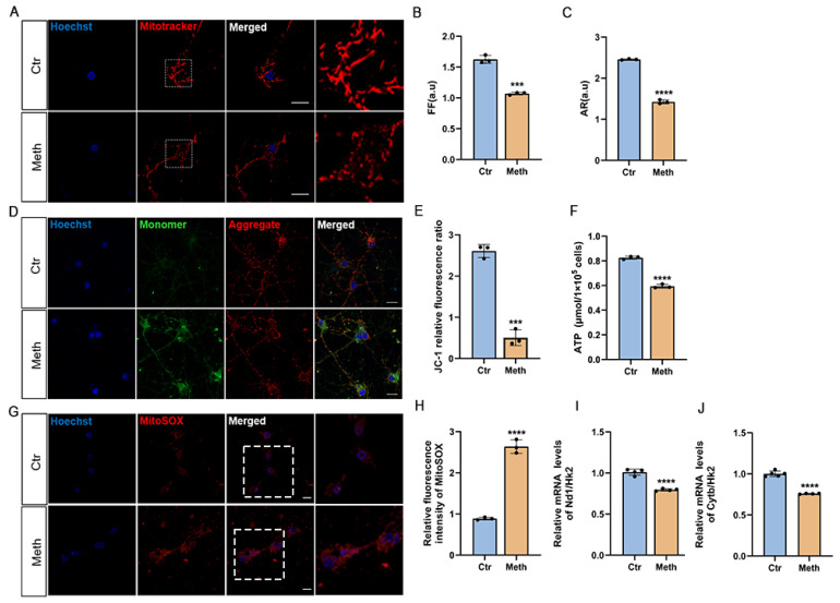
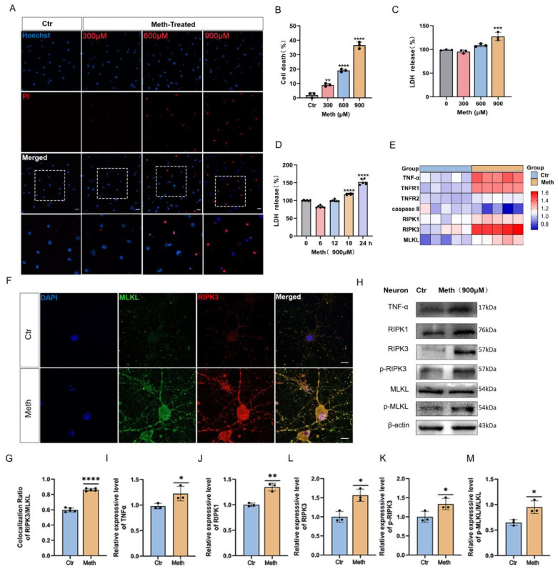
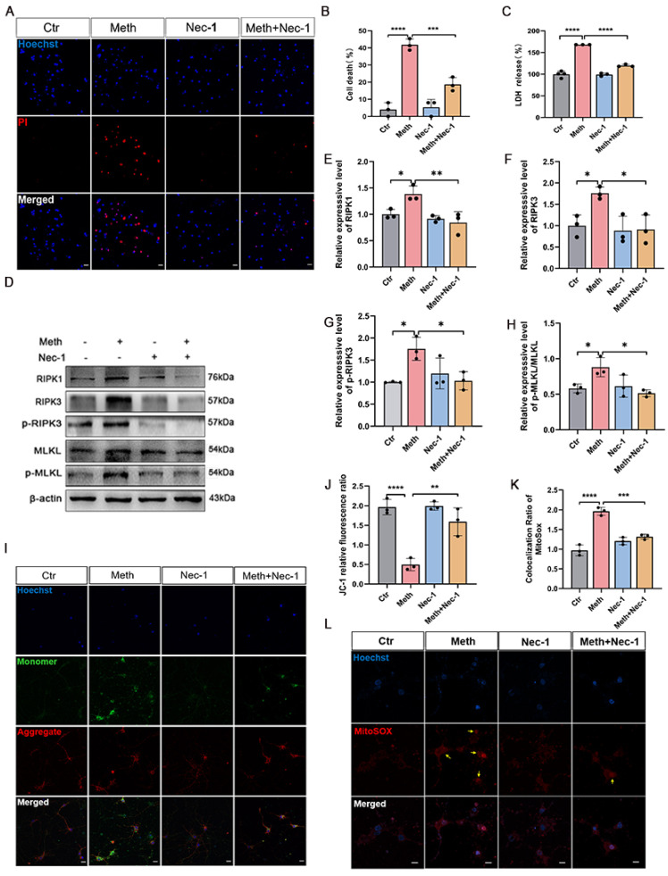
Methamphetamine (Meth), a psychostimulant drug of the amphetamine type, is widely abused and highly neurotoxic. Meth exposure leads to neuronal necroptosis, and the mitochondrial dysfunction may be involved. However, the underlying mechanisms remain poorly understood. Here, we found that Meth significantly elicited the formation of the RIPK1-RIPK3-MLKL necrosome complex. Intriguingly, the activated MLKL (p-MLKL) translocated to the mitochondrial membrane and displayed pore-forming activity, manifesting as the penetration of MLKL in the cell membranes of the mitochondria, which caused decreased mitochondrial membrane potential, ATP generation, and mitochondrial DNA (mtDNA) and increased mitochondrial ROS (mtROS) generation, which finalized neuronal necroptosis. Notably, MLKL activation and translocation seem to depend on the RIPK1-RIPK3 axis since these adverse effects can be substantially ameliorated by disruption of the necrosome complex formation by the necroptotic inhibitor 1 (Nec-1), which also markedly impeded the MLKL mitochondrial membrane translocation. Finally, to delineate the effects of pore formation-associated ROS generation, specific blockage of mtROS retarded the Meth-induced neuronal necroptosis. In conclusion, our study reveals for the first time that MLKL mitochondrial membrane translocation may be involved in Meth-induced neuronal necroptosis. Therefore, impeding MLKL translocation might provide a novel therapeutic strategy for Meth-induced neurotoxicity.
ToxicsChemical Engineering-Chemical Health and Safety
CiteScore
4.50
自引率
10.90%
发文量
681
审稿时长
6 weeks
期刊介绍:
Toxics (ISSN 2305-6304) is an international, peer-reviewed, open access journal which provides an advanced forum for studies related to all aspects of toxic chemicals and materials. It publishes reviews, regular research papers, and short communications. Our aim is to encourage scientists to publish their experimental and theoretical results in detail. There is, therefore, no restriction on the maximum length of the papers, although authors should write their papers in a clear and concise way. The full experimental details must be provided so that the results can be reproduced. Electronic files or software regarding the full details of calculations and experimental procedure can be deposited as supplementary material, if it is not possible to publish them along with the text.
 求助内容:
求助内容: 应助结果提醒方式:


