{"title":"BPAF对海洋Medaka (Oryzias melastigma)胚胎-幼虫期的毒性作用","authors":"Jiahao Gao, Tianyang Zhou, Zuchun Chen, Ning Zhang, Yusong Guo, Zhongduo Wang, Wenjun Shi, Zhongdian Dong","doi":"10.3390/toxics13090773","DOIUrl":null,"url":null,"abstract":"<p><p>BPAF (Bisphenol AF), one of the primary substitutes for BPA (Bisphenol A), is widely used in the production of plastics, optical fibers, and other materials. During the use of these products, BPAF inevitably enters the environment and exerts toxic effects on animal growth, development, reproduction, immunity, neurology, and genetics. This study employed marine medaka (<i>Oryzias melastigma</i>) as the experimental model to evaluate the toxicological impacts of BPAF on early development. Embryos were exposed to four BPAF concentrations (0, 1 μg/L, 10 μg/L, and 100 μg/L) for 14 days (embryonic to larval stages), followed by phenotypic measurements, behavioral analysis, and gene expression detection. The results demonstrated that BPAF exposure induced developmental malformations and reduced survival rates in marine medaka embryos, with embryo survival negatively correlated with BPAF concentrations. Additionally, BPAF significantly decreased embryonic heart rates, and the 100 μg/L BPAF group exhibited prolonged embryo hatching time and reduced hatching success. In newly hatched larvae, BPAF exposure led to decreased body length, reduced heart rates, and significant suppression of swimming activity, characterized by increased resting time and reduced swimming distance. BPAF exposure altered the expression levels of genes associated with cardiovascular function (e.g., <i>tbx2b</i>, <i>arnt2</i>), the HPT axis (e.g., <i>tg</i>, <i>dio3a</i>, <i>trh</i>, <i>trhr2</i>, <i>tpo</i>), and neurodevelopment (e.g., <i>ache</i>, <i>elavl3</i>, <i>gfap</i>) in the medaka larvae. These transcriptional perturbations are proposed as potential molecular mechanisms underlying the observed phenotypic effects, including reduced heart rates and suppressed swimming behavior in the study. Molecularly, BPAF exposure significantly disrupted the expression of genes related to the cardiovascular system, HPT axis, and nervous system.</p>","PeriodicalId":23195,"journal":{"name":"Toxics","volume":"13 9","pages":""},"PeriodicalIF":4.1000,"publicationDate":"2025-09-12","publicationTypes":"Journal Article","fieldsOfStudy":null,"isOpenAccess":false,"openAccessPdf":"https://www.ncbi.nlm.nih.gov/pmc/articles/PMC12473650/pdf/","citationCount":"0","resultStr":"{\"title\":\"Toxic Effects of BPAF on Marine Medaka (<i>Oryzias melastigma</i>) During Embryo-Larval Stages.\",\"authors\":\"Jiahao Gao, Tianyang Zhou, Zuchun Chen, Ning Zhang, Yusong Guo, Zhongduo Wang, Wenjun Shi, Zhongdian Dong\",\"doi\":\"10.3390/toxics13090773\",\"DOIUrl\":null,\"url\":null,\"abstract\":\"<p><p>BPAF (Bisphenol AF), one of the primary substitutes for BPA (Bisphenol A), is widely used in the production of plastics, optical fibers, and other materials. During the use of these products, BPAF inevitably enters the environment and exerts toxic effects on animal growth, development, reproduction, immunity, neurology, and genetics. This study employed marine medaka (<i>Oryzias melastigma</i>) as the experimental model to evaluate the toxicological impacts of BPAF on early development. Embryos were exposed to four BPAF concentrations (0, 1 μg/L, 10 μg/L, and 100 μg/L) for 14 days (embryonic to larval stages), followed by phenotypic measurements, behavioral analysis, and gene expression detection. The results demonstrated that BPAF exposure induced developmental malformations and reduced survival rates in marine medaka embryos, with embryo survival negatively correlated with BPAF concentrations. Additionally, BPAF significantly decreased embryonic heart rates, and the 100 μg/L BPAF group exhibited prolonged embryo hatching time and reduced hatching success. In newly hatched larvae, BPAF exposure led to decreased body length, reduced heart rates, and significant suppression of swimming activity, characterized by increased resting time and reduced swimming distance. BPAF exposure altered the expression levels of genes associated with cardiovascular function (e.g., <i>tbx2b</i>, <i>arnt2</i>), the HPT axis (e.g., <i>tg</i>, <i>dio3a</i>, <i>trh</i>, <i>trhr2</i>, <i>tpo</i>), and neurodevelopment (e.g., <i>ache</i>, <i>elavl3</i>, <i>gfap</i>) in the medaka larvae. These transcriptional perturbations are proposed as potential molecular mechanisms underlying the observed phenotypic effects, including reduced heart rates and suppressed swimming behavior in the study. Molecularly, BPAF exposure significantly disrupted the expression of genes related to the cardiovascular system, HPT axis, and nervous system.</p>\",\"PeriodicalId\":23195,\"journal\":{\"name\":\"Toxics\",\"volume\":\"13 9\",\"pages\":\"\"},\"PeriodicalIF\":4.1000,\"publicationDate\":\"2025-09-12\",\"publicationTypes\":\"Journal Article\",\"fieldsOfStudy\":null,\"isOpenAccess\":false,\"openAccessPdf\":\"https://www.ncbi.nlm.nih.gov/pmc/articles/PMC12473650/pdf/\",\"citationCount\":\"0\",\"resultStr\":null,\"platform\":\"Semanticscholar\",\"paperid\":null,\"PeriodicalName\":\"Toxics\",\"FirstCategoryId\":\"93\",\"ListUrlMain\":\"https://doi.org/10.3390/toxics13090773\",\"RegionNum\":3,\"RegionCategory\":\"环境科学与生态学\",\"ArticlePicture\":[],\"TitleCN\":null,\"AbstractTextCN\":null,\"PMCID\":null,\"EPubDate\":\"\",\"PubModel\":\"\",\"JCR\":\"Q2\",\"JCRName\":\"ENVIRONMENTAL SCIENCES\",\"Score\":null,\"Total\":0}","platform":"Semanticscholar","paperid":null,"PeriodicalName":"Toxics","FirstCategoryId":"93","ListUrlMain":"https://doi.org/10.3390/toxics13090773","RegionNum":3,"RegionCategory":"环境科学与生态学","ArticlePicture":[],"TitleCN":null,"AbstractTextCN":null,"PMCID":null,"EPubDate":"","PubModel":"","JCR":"Q2","JCRName":"ENVIRONMENTAL SCIENCES","Score":null,"Total":0}
Toxic Effects of BPAF on Marine Medaka (Oryzias melastigma) During Embryo-Larval Stages.
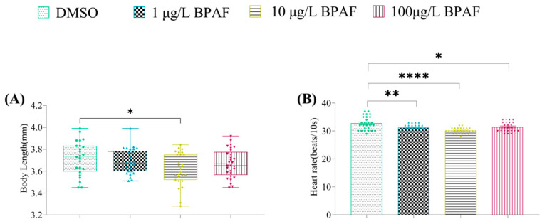
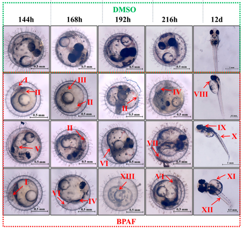
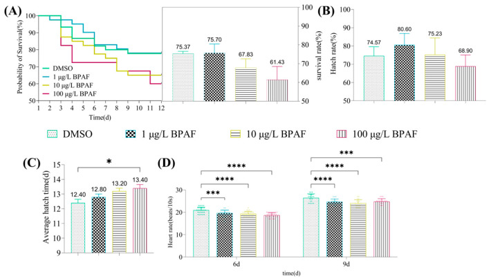
BPAF (Bisphenol AF), one of the primary substitutes for BPA (Bisphenol A), is widely used in the production of plastics, optical fibers, and other materials. During the use of these products, BPAF inevitably enters the environment and exerts toxic effects on animal growth, development, reproduction, immunity, neurology, and genetics. This study employed marine medaka (Oryzias melastigma) as the experimental model to evaluate the toxicological impacts of BPAF on early development. Embryos were exposed to four BPAF concentrations (0, 1 μg/L, 10 μg/L, and 100 μg/L) for 14 days (embryonic to larval stages), followed by phenotypic measurements, behavioral analysis, and gene expression detection. The results demonstrated that BPAF exposure induced developmental malformations and reduced survival rates in marine medaka embryos, with embryo survival negatively correlated with BPAF concentrations. Additionally, BPAF significantly decreased embryonic heart rates, and the 100 μg/L BPAF group exhibited prolonged embryo hatching time and reduced hatching success. In newly hatched larvae, BPAF exposure led to decreased body length, reduced heart rates, and significant suppression of swimming activity, characterized by increased resting time and reduced swimming distance. BPAF exposure altered the expression levels of genes associated with cardiovascular function (e.g., tbx2b, arnt2), the HPT axis (e.g., tg, dio3a, trh, trhr2, tpo), and neurodevelopment (e.g., ache, elavl3, gfap) in the medaka larvae. These transcriptional perturbations are proposed as potential molecular mechanisms underlying the observed phenotypic effects, including reduced heart rates and suppressed swimming behavior in the study. Molecularly, BPAF exposure significantly disrupted the expression of genes related to the cardiovascular system, HPT axis, and nervous system.
ToxicsChemical Engineering-Chemical Health and Safety
CiteScore
4.50
自引率
10.90%
发文量
681
审稿时长
6 weeks
期刊介绍:
Toxics (ISSN 2305-6304) is an international, peer-reviewed, open access journal which provides an advanced forum for studies related to all aspects of toxic chemicals and materials. It publishes reviews, regular research papers, and short communications. Our aim is to encourage scientists to publish their experimental and theoretical results in detail. There is, therefore, no restriction on the maximum length of the papers, although authors should write their papers in a clear and concise way. The full experimental details must be provided so that the results can be reproduced. Electronic files or software regarding the full details of calculations and experimental procedure can be deposited as supplementary material, if it is not possible to publish them along with the text.
 求助内容:
求助内容: 应助结果提醒方式:


