{"title":"分数各向异性作为大鼠缺血再灌注损伤模型血脑屏障破坏的潜在标志","authors":"Duen-Pang Kuo, Hua-Shan Liu, Yung-Chieh Chen, Chen-Yin Ou, Cheng-Yu Chen","doi":"10.1007/s00723-025-01759-y","DOIUrl":null,"url":null,"abstract":"<div><p>While gadolinium-enhanced magnetic resonance imaging (MRI) enables assessment of blood–brain barrier (BBB) integrity post-stroke, it presents inherent risks and limitations. We evaluated diffusion tensor imaging (DTI) as a contrast-free method for monitoring BBB integrity during ischemia–reperfusion (I/R) in a rat stroke model. Seventeen Sprague–Dawley rats underwent one-hour transient middle cerebral artery occlusion (MCAO). Serial MRI sequences, including DTI, perfusion-weighted imaging, and T1 mapping, were performed to monitor BBB permeability (BBBP) dynamics in regions initially identified as ischemic penumbra (IP) and ischemic core (IC) during MCAO. The fractional anisotropy (FA) ratio (rFA) was compared with BBBP changes throughout the observation period. In the IC, rFA values exhibited a 13% increase during ischemia, followed by a 10% decrease post-recanalization, and a 36% decline at 120 h post-reperfusion. Although BBBP in the IC decreased during ischemia and normalized post-reperfusion, persistent leakage was observed. The IP maintained stable rFA and BBB integrity throughout I/R. Strong negative correlations between rFA and BBBP were found in ischemic regions (IC: r = − 0.90, p < 0.001; IP: r = − 0.78, p = 0.007), with FA reductions at 24 h post-reperfusion predicting BBB disruption at 120 h. These findings suggest that DTI metrics correlate significantly with BBB integrity during I/R, indicating potential utility as a contrast-free monitoring tool for early detection of BBB impairment following stroke.</p></div>","PeriodicalId":469,"journal":{"name":"Applied Magnetic Resonance","volume":"56 7","pages":"845 - 858"},"PeriodicalIF":1.1000,"publicationDate":"2025-04-23","publicationTypes":"Journal Article","fieldsOfStudy":null,"isOpenAccess":false,"openAccessPdf":"","citationCount":"0","resultStr":"{\"title\":\"Fractional Anisotropy as a Potential Marker of Blood–Brain Barrier Disruption in a Rat Model of Ischemia–Reperfusion Injury\",\"authors\":\"Duen-Pang Kuo, Hua-Shan Liu, Yung-Chieh Chen, Chen-Yin Ou, Cheng-Yu Chen\",\"doi\":\"10.1007/s00723-025-01759-y\",\"DOIUrl\":null,\"url\":null,\"abstract\":\"<div><p>While gadolinium-enhanced magnetic resonance imaging (MRI) enables assessment of blood–brain barrier (BBB) integrity post-stroke, it presents inherent risks and limitations. We evaluated diffusion tensor imaging (DTI) as a contrast-free method for monitoring BBB integrity during ischemia–reperfusion (I/R) in a rat stroke model. Seventeen Sprague–Dawley rats underwent one-hour transient middle cerebral artery occlusion (MCAO). Serial MRI sequences, including DTI, perfusion-weighted imaging, and T1 mapping, were performed to monitor BBB permeability (BBBP) dynamics in regions initially identified as ischemic penumbra (IP) and ischemic core (IC) during MCAO. The fractional anisotropy (FA) ratio (rFA) was compared with BBBP changes throughout the observation period. In the IC, rFA values exhibited a 13% increase during ischemia, followed by a 10% decrease post-recanalization, and a 36% decline at 120 h post-reperfusion. Although BBBP in the IC decreased during ischemia and normalized post-reperfusion, persistent leakage was observed. The IP maintained stable rFA and BBB integrity throughout I/R. Strong negative correlations between rFA and BBBP were found in ischemic regions (IC: r = − 0.90, p < 0.001; IP: r = − 0.78, p = 0.007), with FA reductions at 24 h post-reperfusion predicting BBB disruption at 120 h. These findings suggest that DTI metrics correlate significantly with BBB integrity during I/R, indicating potential utility as a contrast-free monitoring tool for early detection of BBB impairment following stroke.</p></div>\",\"PeriodicalId\":469,\"journal\":{\"name\":\"Applied Magnetic Resonance\",\"volume\":\"56 7\",\"pages\":\"845 - 858\"},\"PeriodicalIF\":1.1000,\"publicationDate\":\"2025-04-23\",\"publicationTypes\":\"Journal Article\",\"fieldsOfStudy\":null,\"isOpenAccess\":false,\"openAccessPdf\":\"\",\"citationCount\":\"0\",\"resultStr\":null,\"platform\":\"Semanticscholar\",\"paperid\":null,\"PeriodicalName\":\"Applied Magnetic Resonance\",\"FirstCategoryId\":\"101\",\"ListUrlMain\":\"https://link.springer.com/article/10.1007/s00723-025-01759-y\",\"RegionNum\":4,\"RegionCategory\":\"物理与天体物理\",\"ArticlePicture\":[],\"TitleCN\":null,\"AbstractTextCN\":null,\"PMCID\":null,\"EPubDate\":\"\",\"PubModel\":\"\",\"JCR\":\"Q4\",\"JCRName\":\"PHYSICS, ATOMIC, MOLECULAR & CHEMICAL\",\"Score\":null,\"Total\":0}","platform":"Semanticscholar","paperid":null,"PeriodicalName":"Applied Magnetic Resonance","FirstCategoryId":"101","ListUrlMain":"https://link.springer.com/article/10.1007/s00723-025-01759-y","RegionNum":4,"RegionCategory":"物理与天体物理","ArticlePicture":[],"TitleCN":null,"AbstractTextCN":null,"PMCID":null,"EPubDate":"","PubModel":"","JCR":"Q4","JCRName":"PHYSICS, ATOMIC, MOLECULAR & CHEMICAL","Score":null,"Total":0}
引用次数: 0
摘要
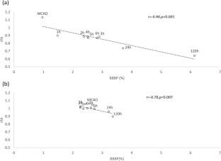
虽然钆增强磁共振成像(MRI)能够评估脑卒中后血脑屏障(BBB)的完整性,但它存在固有的风险和局限性。我们评估了弥散张量成像(DTI)作为无造影剂监测大鼠脑卒中模型缺血再灌注(I/R)期间血脑屏障完整性的方法。17只Sprague-Dawley大鼠进行1小时短暂性大脑中动脉闭塞(MCAO)。连续MRI序列,包括DTI、灌注加权成像和T1定位,监测MCAO期间最初被确定为缺血半暗带(IP)和缺血核心(IC)区域的血脑屏障通透性(BBBP)动态。各向异性分数(FA)比(rFA)与BBBP在整个观察期内的变化进行比较。在IC中,rFA值在缺血期间增加13%,再通后下降10%,再灌注后120 h下降36%。虽然在缺血和再灌注后恢复正常时,IC中的BBBP降低,但仍观察到持续的泄漏。在整个I/R过程中,IP保持稳定的rFA和血脑屏障完整性。在缺血区域,rFA和BBBP之间存在很强的负相关(IC: r = - 0.90, p < 0.001; IP: r = - 0.78, p = 0.007),再灌注后24小时的FA减少预测120小时的血脑屏障破坏。这些发现表明,DTI指标与I/ r期间血脑屏障完整性显著相关,表明作为卒中后血脑屏障损伤早期检测的无对比监测工具的潜在应用价值。
Fractional Anisotropy as a Potential Marker of Blood–Brain Barrier Disruption in a Rat Model of Ischemia–Reperfusion Injury
While gadolinium-enhanced magnetic resonance imaging (MRI) enables assessment of blood–brain barrier (BBB) integrity post-stroke, it presents inherent risks and limitations. We evaluated diffusion tensor imaging (DTI) as a contrast-free method for monitoring BBB integrity during ischemia–reperfusion (I/R) in a rat stroke model. Seventeen Sprague–Dawley rats underwent one-hour transient middle cerebral artery occlusion (MCAO). Serial MRI sequences, including DTI, perfusion-weighted imaging, and T1 mapping, were performed to monitor BBB permeability (BBBP) dynamics in regions initially identified as ischemic penumbra (IP) and ischemic core (IC) during MCAO. The fractional anisotropy (FA) ratio (rFA) was compared with BBBP changes throughout the observation period. In the IC, rFA values exhibited a 13% increase during ischemia, followed by a 10% decrease post-recanalization, and a 36% decline at 120 h post-reperfusion. Although BBBP in the IC decreased during ischemia and normalized post-reperfusion, persistent leakage was observed. The IP maintained stable rFA and BBB integrity throughout I/R. Strong negative correlations between rFA and BBBP were found in ischemic regions (IC: r = − 0.90, p < 0.001; IP: r = − 0.78, p = 0.007), with FA reductions at 24 h post-reperfusion predicting BBB disruption at 120 h. These findings suggest that DTI metrics correlate significantly with BBB integrity during I/R, indicating potential utility as a contrast-free monitoring tool for early detection of BBB impairment following stroke.
期刊介绍:
Applied Magnetic Resonance provides an international forum for the application of magnetic resonance in physics, chemistry, biology, medicine, geochemistry, ecology, engineering, and related fields.
The contents include articles with a strong emphasis on new applications, and on new experimental methods. Additional features include book reviews and Letters to the Editor.
 求助内容:
求助内容: 应助结果提醒方式:


