Bhaurao R. Balbudhe, Dilip S. Badwaik, Shrikant M. Suryawanshi, Sarang R. Daf, Atul N. Yerpude
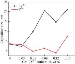
{"title":"Ce3+和Y3+稀土添加对Mn-Zn尖晶石纳米铁素体结构、光学、形貌和磁性能的影响","authors":"Bhaurao R. Balbudhe, Dilip S. Badwaik, Shrikant M. Suryawanshi, Sarang R. Daf, Atul N. Yerpude","doi":"10.1134/S1063783425600517","DOIUrl":null,"url":null,"abstract":"<p>Two series of Mn<sub>0.5</sub>Zn<sub>0.5</sub>Fe<sub>2–<i>x</i></sub>O<sub>4</sub>R<sub><i>x</i></sub> (where R = Ce, Y, and <i>x</i> = 0.00 to 0.15) spinel nanoferrites were synthesized via a co-precipitation approach. Methods including X-ray diffraction (XRD), Fourier transform infrared spectroscopy (FTIR), vibrating sample magnetometer (VSM), and scanning electron microscopy (SEM) were utilized to examine the samples’ structural, morphological, optical, and magnetic features. XRD confirmed a cubic spinel structure, with crystalline sizes lies between 16 and 24 nm for Ce<sup>3+</sup> added and 15 and 19 nm for Y<sup>3+</sup> added ferrite NPs. XRD analysis showed that Ce<sup>3+</sup> and Y<sup>3+</sup> ions were successfully incorporated into the Mn–Zn spinel structure. FTIR spectra validated the presence of tetrahedral (A) and octahedral (B) sites in all compositions of Mn<sub>0.5</sub>Zn<sub>0.5</sub>Fe<sub>2–<i>x</i></sub>O<sub>4</sub>R<sub><i>x</i></sub> nanoparticles, indicative of spinel ferrites exhibiting a face-centered cubic (FCC) structure. SEM studies revealed agglomerated nanoparticles with spherical morphology. Energy dispersive X-ray spectroscopy (EDS) verified that all elements are present in the composition. The TEM micrograph shows the existence of slightly agglomerated nanoparticles. Magnetic properties, including saturation magnetization and coercivity, were analyzed using M–H hysteresis curves, showing dependence on rare earth substitution and A–B exchange interactions. The lower value of coercivity (<i>H</i><sub><i>c</i></sub>) indicatied of soft nature of NPs. The multidomain nature of the nanoferrites indicates their potential for electronics applications.</p>","PeriodicalId":731,"journal":{"name":"Physics of the Solid State","volume":"67 6","pages":"485 - 498"},"PeriodicalIF":1.8000,"publicationDate":"2025-06-18","publicationTypes":"Journal Article","fieldsOfStudy":null,"isOpenAccess":false,"openAccessPdf":"","citationCount":"0","resultStr":"{\"title\":\"Impact of Ce3+ and Y3+ Rare Earth Additions on Structural, Optical, Morphological, and Magnetic Properties of Mn–Zn Spinel Nanoferrites\",\"authors\":\"Bhaurao R. Balbudhe, Dilip S. Badwaik, Shrikant M. Suryawanshi, Sarang R. Daf, Atul N. Yerpude\",\"doi\":\"10.1134/S1063783425600517\",\"DOIUrl\":null,\"url\":null,\"abstract\":\"<p>Two series of Mn<sub>0.5</sub>Zn<sub>0.5</sub>Fe<sub>2–<i>x</i></sub>O<sub>4</sub>R<sub><i>x</i></sub> (where R = Ce, Y, and <i>x</i> = 0.00 to 0.15) spinel nanoferrites were synthesized via a co-precipitation approach. Methods including X-ray diffraction (XRD), Fourier transform infrared spectroscopy (FTIR), vibrating sample magnetometer (VSM), and scanning electron microscopy (SEM) were utilized to examine the samples’ structural, morphological, optical, and magnetic features. XRD confirmed a cubic spinel structure, with crystalline sizes lies between 16 and 24 nm for Ce<sup>3+</sup> added and 15 and 19 nm for Y<sup>3+</sup> added ferrite NPs. XRD analysis showed that Ce<sup>3+</sup> and Y<sup>3+</sup> ions were successfully incorporated into the Mn–Zn spinel structure. FTIR spectra validated the presence of tetrahedral (A) and octahedral (B) sites in all compositions of Mn<sub>0.5</sub>Zn<sub>0.5</sub>Fe<sub>2–<i>x</i></sub>O<sub>4</sub>R<sub><i>x</i></sub> nanoparticles, indicative of spinel ferrites exhibiting a face-centered cubic (FCC) structure. SEM studies revealed agglomerated nanoparticles with spherical morphology. Energy dispersive X-ray spectroscopy (EDS) verified that all elements are present in the composition. The TEM micrograph shows the existence of slightly agglomerated nanoparticles. Magnetic properties, including saturation magnetization and coercivity, were analyzed using M–H hysteresis curves, showing dependence on rare earth substitution and A–B exchange interactions. The lower value of coercivity (<i>H</i><sub><i>c</i></sub>) indicatied of soft nature of NPs. The multidomain nature of the nanoferrites indicates their potential for electronics applications.</p>\",\"PeriodicalId\":731,\"journal\":{\"name\":\"Physics of the Solid State\",\"volume\":\"67 6\",\"pages\":\"485 - 498\"},\"PeriodicalIF\":1.8000,\"publicationDate\":\"2025-06-18\",\"publicationTypes\":\"Journal Article\",\"fieldsOfStudy\":null,\"isOpenAccess\":false,\"openAccessPdf\":\"\",\"citationCount\":\"0\",\"resultStr\":null,\"platform\":\"Semanticscholar\",\"paperid\":null,\"PeriodicalName\":\"Physics of the Solid State\",\"FirstCategoryId\":\"101\",\"ListUrlMain\":\"https://link.springer.com/article/10.1134/S1063783425600517\",\"RegionNum\":4,\"RegionCategory\":\"物理与天体物理\",\"ArticlePicture\":[],\"TitleCN\":null,\"AbstractTextCN\":null,\"PMCID\":null,\"EPubDate\":\"\",\"PubModel\":\"\",\"JCR\":\"Q4\",\"JCRName\":\"PHYSICS, CONDENSED MATTER\",\"Score\":null,\"Total\":0}","platform":"Semanticscholar","paperid":null,"PeriodicalName":"Physics of the Solid State","FirstCategoryId":"101","ListUrlMain":"https://link.springer.com/article/10.1134/S1063783425600517","RegionNum":4,"RegionCategory":"物理与天体物理","ArticlePicture":[],"TitleCN":null,"AbstractTextCN":null,"PMCID":null,"EPubDate":"","PubModel":"","JCR":"Q4","JCRName":"PHYSICS, CONDENSED MATTER","Score":null,"Total":0}
引用次数: 0
摘要
采用共沉淀法合成了两系Mn0.5Zn0.5Fe2-xO4Rx (R = Ce, Y, x = 0.00 ~ 0.15)尖晶石纳米铁素体。利用x射线衍射(XRD)、傅里叶变换红外光谱(FTIR)、振动样品磁强计(VSM)和扫描电镜(SEM)等方法对样品的结构、形态、光学和磁性进行了表征。XRD证实为立方尖晶石结构,添加Ce3+的铁素体NPs晶粒尺寸在16 ~ 24 nm之间,添加Y3+的铁素体NPs晶粒尺寸在15 ~ 19 nm之间。XRD分析表明,Ce3+和Y3+离子成功地掺入到Mn-Zn尖晶石结构中。FTIR光谱验证了Mn0.5Zn0.5Fe2-xO4Rx纳米颗粒的组成中存在四面体(A)和八面体(B)位点,表明尖晶石铁氧体具有面心立方(FCC)结构。扫描电镜研究表明,纳米颗粒凝聚成球状。能量色散x射线光谱(EDS)证实了所有元素都存在于组成中。TEM显微图显示存在微团聚的纳米颗粒。利用M-H磁滞曲线分析磁性能,包括饱和磁化和矫顽力,显示稀土取代和A-B交换相互作用的依赖性。较低的矫顽力(Hc)值表明NPs的软性质。纳米铁氧体的多畴特性表明了它们在电子领域的应用潜力。
Impact of Ce3+ and Y3+ Rare Earth Additions on Structural, Optical, Morphological, and Magnetic Properties of Mn–Zn Spinel Nanoferrites
Two series of Mn0.5Zn0.5Fe2–xO4Rx (where R = Ce, Y, and x = 0.00 to 0.15) spinel nanoferrites were synthesized via a co-precipitation approach. Methods including X-ray diffraction (XRD), Fourier transform infrared spectroscopy (FTIR), vibrating sample magnetometer (VSM), and scanning electron microscopy (SEM) were utilized to examine the samples’ structural, morphological, optical, and magnetic features. XRD confirmed a cubic spinel structure, with crystalline sizes lies between 16 and 24 nm for Ce3+ added and 15 and 19 nm for Y3+ added ferrite NPs. XRD analysis showed that Ce3+ and Y3+ ions were successfully incorporated into the Mn–Zn spinel structure. FTIR spectra validated the presence of tetrahedral (A) and octahedral (B) sites in all compositions of Mn0.5Zn0.5Fe2–xO4Rx nanoparticles, indicative of spinel ferrites exhibiting a face-centered cubic (FCC) structure. SEM studies revealed agglomerated nanoparticles with spherical morphology. Energy dispersive X-ray spectroscopy (EDS) verified that all elements are present in the composition. The TEM micrograph shows the existence of slightly agglomerated nanoparticles. Magnetic properties, including saturation magnetization and coercivity, were analyzed using M–H hysteresis curves, showing dependence on rare earth substitution and A–B exchange interactions. The lower value of coercivity (Hc) indicatied of soft nature of NPs. The multidomain nature of the nanoferrites indicates their potential for electronics applications.
期刊介绍:
Presents the latest results from Russia’s leading researchers in condensed matter physics at the Russian Academy of Sciences and other prestigious institutions. Covers all areas of solid state physics including solid state optics, solid state acoustics, electronic and vibrational spectra, phase transitions, ferroelectricity, magnetism, and superconductivity. Also presents review papers on the most important problems in solid state physics.
 求助内容:
求助内容: 应助结果提醒方式:


