用于光子计数CT图像展开的胰管中心线提取
IF 2.8
4区 计算机科学
Q2 COMPUTER SCIENCE, SOFTWARE ENGINEERING
引用次数: 0
摘要
胰腺疾病通常在晚期才被诊断出来,由于死亡率非常高,胰腺癌是最令人恐惧的。主胰管的异常,如阻塞和扩张,通常是这类胰腺疾病的(早期)征象,但在标准的计算机断层扫描图像系列中很难发现。光子计数计算机断层扫描具有更高的分辨率,提高了该导管的可检测性,允许诊断评估。单一视图的全面可视化需要以中心线为基础展开导管和胰腺。然而,手工中心线注释是乏味的。为了使这一过程自动化,我们引入了一个全自动的胰管展开管道,通过使用Dijkstra算法在从分割概率图派生的成本图上鲁棒提取中心线。这项工作的核心贡献在于处理数据驱动的成本图,从而为生成胰腺的CPR可视化提供一致的中心线。为了改进管道中的各个步骤,我们进一步研究了分割滤波和拓扑保持骨架召回损失等增强功能。在评估中,我们评估了我们的方法在超高分辨率和常规PCCT图像上的性能。我们发现,从两种扫描类型中都可以一致地提取中心线,其中超高分辨率图像的中心线的中位数误差为0.58 mm,而使用常规分辨率的中位数误差为0.73 mm。本文章由计算机程序翻译,如有差异,请以英文原文为准。
Pancreatic duct centerline extraction for image unfolding in photon-counting CT
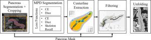
Pancreatic diseases are often only diagnosed at a late stage, and pancreatic cancer is the most feared due to a very high mortality. Abnormalities of the main pancreatic duct, such as blockages and dilatation, are often (early) signs of such pancreatic diseases, but are difficult to detect in standard Computed Tomography image series. Photon-Counting Computed Tomography with its higher resolution improves the detectability of this duct, allowing diagnostic assessment. A comprehensive visualization in a single view requires a centerline-based unfolding of the duct and pancreas. However, manual centerline annotation is tedious. To automate this process, we introduce a fully automated pipeline for pancreatic duct unfolding by robustly extracting the centerline using Dijkstra’s algorithm on a cost map derived from a segmentation probability map. The core contribution of this work lies in the processing of the data-driven cost map leading to a consistent centerline for generating CPR visualizations of the pancreas. To improve individual steps within the pipeline, we investigate further enhancements such as segmentation filtering and the topology-preserving skeleton recall loss. In the evaluation, we assess performance of our method on both ultra-high-resolution and regular PCCT images. We find that the centerline can be consistently extracted from both scan types, where the centerlines from the ultra-high resolution images exhibit a slightly lower median error of 0.58 mm compared to the 0.73 mm using the regular resolution.
求助全文
通过发布文献求助,成功后即可免费获取论文全文。
去求助
来源期刊
Computers & Graphics-Uk
工程技术-计算机:软件工程
CiteScore
5.30
自引率
12.00%
发文量
173
审稿时长
38 days
期刊介绍:
Computers & Graphics is dedicated to disseminate information on research and applications of computer graphics (CG) techniques. The journal encourages articles on:
1. Research and applications of interactive computer graphics. We are particularly interested in novel interaction techniques and applications of CG to problem domains.
2. State-of-the-art papers on late-breaking, cutting-edge research on CG.
3. Information on innovative uses of graphics principles and technologies.
4. Tutorial papers on both teaching CG principles and innovative uses of CG in education.
 求助内容:
求助内容: 应助结果提醒方式:


