基于人工智能的外科解剖可视化教学效果:一项聚类准随机对照试验
IF 2.7
2区 医学
Q1 SURGERY
引用次数: 0
摘要
尽管外科技术有了显著的进步,但医学生的外科教育在很大程度上仍然是传统的。由于准确识别术中解剖结构的重要性,对于新手来说,被动观察是不够的。本研究评估了基于人工智能的可视化系统的教育效果,该系统在手术过程中实时突出解剖结构。方法对五年级医学生进行整群准随机对照试验,两组分别接受常规术中教学(队列C)和人工智能可视化辅助教学(队列a)。每位学生在临床观察训练前后分别在6张静态腹腔镜或机器人图像上标注胰腺区域。使用3个图像分割指标评估性能:召回率(最小化假阴性),精度(避免假阳性)和Dice系数(集成召回率和精度)。Dice系数是主要结果,召回率和准确率是次要结果。结果两组患者的基线表现相似,训练后所有指标均有所改善。然而,队列A在回忆率方面的改善明显大于队列C (ΔRecall: 0.112 vs 0.013, P = 0.020),表明识别胰腺区域的敏感性增强。两组间精密度无显著差异(P = .746)。A组的Dice系数比C组改善更多(ΔDice: 0.087 vs 0.028),但组间差异无统计学意义(P = 0.097)。散点图分析显示,许多队列A学生在不损失精度的情况下,记忆力有所提高,表明识别精度有所提高。结论将人工智能可视化技术应用于外科解剖教学中,可提高医学生对术中解剖的认识,特别是对胰腺等视觉复杂结构的认识。本文章由计算机程序翻译,如有差异,请以英文原文为准。
Effectiveness of artificial intelligence–based visualization for surgical anatomy education: A cluster quasirandomized controlled trial
Background
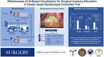
Although surgical techniques have advanced markedly, surgical education for medical students has remained largely traditional. Because of the importance of accurate recognition of intraoperative anatomy, passive observation is insufficient for novice learners. This study evaluated the educational effectiveness of an artificial intelligence–based visualization system that highlights anatomical structures during surgery in real time.
Methods
We conducted a cluster quasi-randomized controlled trial involving fifth-year medical students assigned to receive either conventional intraoperative teaching (cohort C) or instruction augmented by artificial intelligence–based visualization (cohort A). Each student annotated the pancreatic regions on 6 static laparoscopic or robotic images before and after clinical observation training. Performance was assessed using 3 image segmentation metrics: recall (minimizing false negatives), precision (avoiding false positives), and the Dice coefficient, which integrates recall and precision. The Dice coefficient served as the primary outcome, whereas recall and precision served as secondary outcomes.
Results
Both cohorts showed similar baseline performance and improved across all metrics after training. However, cohort A exhibited a significantly greater improvement in recall than cohort C (ΔRecall: 0.112 vs 0.013, P = .020), indicating enhanced sensitivity in identifying pancreatic regions. Precision did not differ significantly between the groups (P = .746). The Dice coefficient improved more in cohort A than in cohort C (ΔDice: 0.087 vs 0.028), although the between-group difference did not reach statistical significance (P = .097). Scatter plot analysis showed that many cohort A students exhibited increased recall without loss of precision, suggesting improved recognition accuracy.
Conclusion
Incorporating artificial intelligence–based visualization into surgical anatomy education may enhance medical students' recognition of intraoperative anatomy, particularly for visually complex structures such as the pancreas.
求助全文
通过发布文献求助,成功后即可免费获取论文全文。
去求助
来源期刊
Surgery
医学-外科
CiteScore
5.40
自引率
5.30%
发文量
687
审稿时长
64 days
期刊介绍:
For 66 years, Surgery has published practical, authoritative information about procedures, clinical advances, and major trends shaping general surgery. Each issue features original scientific contributions and clinical reports. Peer-reviewed articles cover topics in oncology, trauma, gastrointestinal, vascular, and transplantation surgery. The journal also publishes papers from the meetings of its sponsoring societies, the Society of University Surgeons, the Central Surgical Association, and the American Association of Endocrine Surgeons.
 求助内容:
求助内容: 应助结果提醒方式:


