William J Tipping, Gwyn W Gould, Karen Faulds, Duncan Graham
{"title":"利用相量分析在拉曼光谱无噪区解混高光谱SRS图像。","authors":"William J Tipping, Gwyn W Gould, Karen Faulds, Duncan Graham","doi":"10.1021/cbmi.5c00023","DOIUrl":null,"url":null,"abstract":"<p><p>Hyperspectral stimulated Raman scattering (SRS) microscopy is rapidly becoming an established method for chemical and biomedical imaging due to the combination of high spatial resolution and chemical information contained within the three-dimensional data set. Chemometric analysis techniques based on linear unmixing, or multivariate analysis, have become indispensable when visualizing hyperspectral data sets. The application of spectral phasor analysis has also been extremely fruitful in this regard, providing a convenient method to retrieve the spatial and chemical components of the data set. Here, we demonstrate the application of spectral phasor analysis for unmixing the overlapping spectral features within the cell-silent region of the SRS spectrum (2000-2300 cm<sup>-1</sup>). In doing so, we show it is possible to identify specific Raman signals for DNA, proteins, and lipids following glucose-d<sub>7</sub> metabolism in dividing cells. In addition, we show that spectral phasor analysis is capable of distinguishing different bioorthogonal Raman signals including alkynes and carbon-deuterium (C-D) bonds. We demonstrate the application of spectral phasor analysis for multicomponent unmixing of bioorthogonal Raman groups for high-content cellular imaging applications.</p>","PeriodicalId":53181,"journal":{"name":"Chemical & Biomedical Imaging","volume":"3 9","pages":"630-635"},"PeriodicalIF":5.7000,"publicationDate":"2025-05-13","publicationTypes":"Journal Article","fieldsOfStudy":null,"isOpenAccess":false,"openAccessPdf":"https://www.ncbi.nlm.nih.gov/pmc/articles/PMC12457996/pdf/","citationCount":"0","resultStr":"{\"title\":\"Unmixing Hyperspectral SRS Images in the Cell-Silent Region of the Raman Spectrum Using Phasor Analysis.\",\"authors\":\"William J Tipping, Gwyn W Gould, Karen Faulds, Duncan Graham\",\"doi\":\"10.1021/cbmi.5c00023\",\"DOIUrl\":null,\"url\":null,\"abstract\":\"<p><p>Hyperspectral stimulated Raman scattering (SRS) microscopy is rapidly becoming an established method for chemical and biomedical imaging due to the combination of high spatial resolution and chemical information contained within the three-dimensional data set. Chemometric analysis techniques based on linear unmixing, or multivariate analysis, have become indispensable when visualizing hyperspectral data sets. The application of spectral phasor analysis has also been extremely fruitful in this regard, providing a convenient method to retrieve the spatial and chemical components of the data set. Here, we demonstrate the application of spectral phasor analysis for unmixing the overlapping spectral features within the cell-silent region of the SRS spectrum (2000-2300 cm<sup>-1</sup>). In doing so, we show it is possible to identify specific Raman signals for DNA, proteins, and lipids following glucose-d<sub>7</sub> metabolism in dividing cells. In addition, we show that spectral phasor analysis is capable of distinguishing different bioorthogonal Raman signals including alkynes and carbon-deuterium (C-D) bonds. We demonstrate the application of spectral phasor analysis for multicomponent unmixing of bioorthogonal Raman groups for high-content cellular imaging applications.</p>\",\"PeriodicalId\":53181,\"journal\":{\"name\":\"Chemical & Biomedical Imaging\",\"volume\":\"3 9\",\"pages\":\"630-635\"},\"PeriodicalIF\":5.7000,\"publicationDate\":\"2025-05-13\",\"publicationTypes\":\"Journal Article\",\"fieldsOfStudy\":null,\"isOpenAccess\":false,\"openAccessPdf\":\"https://www.ncbi.nlm.nih.gov/pmc/articles/PMC12457996/pdf/\",\"citationCount\":\"0\",\"resultStr\":null,\"platform\":\"Semanticscholar\",\"paperid\":null,\"PeriodicalName\":\"Chemical & Biomedical Imaging\",\"FirstCategoryId\":\"1085\",\"ListUrlMain\":\"https://doi.org/10.1021/cbmi.5c00023\",\"RegionNum\":0,\"RegionCategory\":null,\"ArticlePicture\":[],\"TitleCN\":null,\"AbstractTextCN\":null,\"PMCID\":null,\"EPubDate\":\"2025/9/22 0:00:00\",\"PubModel\":\"eCollection\",\"JCR\":\"\",\"JCRName\":\"\",\"Score\":null,\"Total\":0}","platform":"Semanticscholar","paperid":null,"PeriodicalName":"Chemical & Biomedical Imaging","FirstCategoryId":"1085","ListUrlMain":"https://doi.org/10.1021/cbmi.5c00023","RegionNum":0,"RegionCategory":null,"ArticlePicture":[],"TitleCN":null,"AbstractTextCN":null,"PMCID":null,"EPubDate":"2025/9/22 0:00:00","PubModel":"eCollection","JCR":"","JCRName":"","Score":null,"Total":0}
Unmixing Hyperspectral SRS Images in the Cell-Silent Region of the Raman Spectrum Using Phasor Analysis.
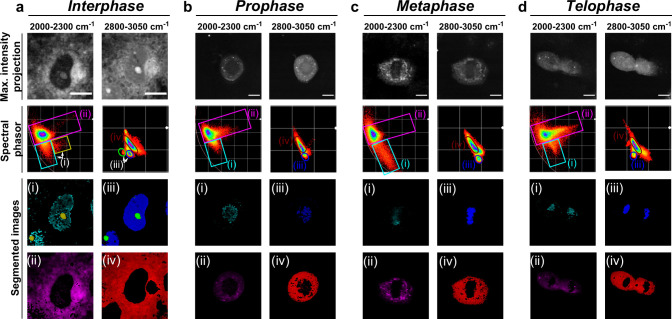
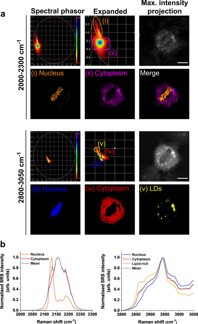
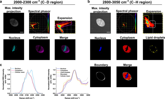
Hyperspectral stimulated Raman scattering (SRS) microscopy is rapidly becoming an established method for chemical and biomedical imaging due to the combination of high spatial resolution and chemical information contained within the three-dimensional data set. Chemometric analysis techniques based on linear unmixing, or multivariate analysis, have become indispensable when visualizing hyperspectral data sets. The application of spectral phasor analysis has also been extremely fruitful in this regard, providing a convenient method to retrieve the spatial and chemical components of the data set. Here, we demonstrate the application of spectral phasor analysis for unmixing the overlapping spectral features within the cell-silent region of the SRS spectrum (2000-2300 cm-1). In doing so, we show it is possible to identify specific Raman signals for DNA, proteins, and lipids following glucose-d7 metabolism in dividing cells. In addition, we show that spectral phasor analysis is capable of distinguishing different bioorthogonal Raman signals including alkynes and carbon-deuterium (C-D) bonds. We demonstrate the application of spectral phasor analysis for multicomponent unmixing of bioorthogonal Raman groups for high-content cellular imaging applications.
期刊介绍:
Chemical & Biomedical Imaging is a peer-reviewed open access journal devoted to the publication of cutting-edge research papers on all aspects of chemical and biomedical imaging. This interdisciplinary field sits at the intersection of chemistry physics biology materials engineering and medicine. The journal aims to bring together researchers from across these disciplines to address cutting-edge challenges of fundamental research and applications.Topics of particular interest include but are not limited to:Imaging of processes and reactionsImaging of nanoscale microscale and mesoscale materialsImaging of biological interactions and interfacesSingle-molecule and cellular imagingWhole-organ and whole-body imagingMolecular imaging probes and contrast agentsBioluminescence chemiluminescence and electrochemiluminescence imagingNanophotonics and imagingChemical tools for new imaging modalitiesChemical and imaging techniques in diagnosis and therapyImaging-guided drug deliveryAI and machine learning assisted imaging
 求助内容:
求助内容: 应助结果提醒方式:


