{"title":"十二指肠尤文氏肉瘤:一种罕见的胃肠道表现:病例报告及文献复习。","authors":"Sujata Agrawal, Paramita Paul","doi":"10.1186/s12957-025-03986-w","DOIUrl":null,"url":null,"abstract":"<p><p>Ewing's sarcoma (ES) is exceptionally rare in the gastrointestinal tract, with only a few cases documented in the literature. We present a case of duodenal Ewing's sarcoma in a 44-year-old woman. The patient presented with severe abdominal pain, vomiting, and loss of appetite. Upper gastrointestinal endoscopy revealed a huge bulge in the first part of the duodenum (D1) wall, resulting in luminal narrowing in D1 and D2. A contrast-enhanced computed tomography scan corroborated the findings with no evidence of any distal metastasis. The patient is a known hypothyroid and had undergone hysterectomy five years back and cholecystectomy one year back. The patient underwent a Whipple procedure along with lymph node dissection. During surgery, a 4.5 cm mass was discovered in the D1 extending to the D2 segment of the duodenum. Histological examination showed a small round cell tumor in the submucosal region. The morphological differentials considered were gastrointestinal stromal tumor (GIST), poorly differentiated carcinoma, ES, neuroendocrine carcinoma (NEC) and lymphoma. Immunohistochemically the tumor cells tested positive for vimentin, NKX2.2, and CD99 (MIC2), and negative for markers of epithelial malignancy (AE1/AE3), lymphoma (CD45), melanoma (HMB45), NEC (INMS1,synaptophysin, chromogranin) and GIST (c-kit). One of the peri-duodenal lymph nodes showed a metastatic deposit. The case was diagnosed as Ewing's sarcoma (extra skeletal), TNM stage pT1 pN1. It was further ratified through fluorescence in situ hybridization (FISH), which showed a EWSR1 (22q12.1) gene rearrangement. A post-surgery PET scan indicated no residual disease. The patient had been receiving post-surgical adjuvant therapy and has completed four cycles of VAC (vincristine dactinomycin cyclophosphamide) and two cycles of VIME (Vincristine, Ifosfamide, Mesna, and Etoposide). The patient is disease-free with one-year follow-up. Follow up with PET scan is being performed at 3 months interval for the first year. The prognosis for Ewing's sarcoma in the gastrointestinal tract is not well-documented; however, a recent review of cases involving the small intestine suggests a poor prognosis. A follow up PET scanning every three months initially would help to target early recurrences and the interval can be increased after 2 years. Also emerging techniques like liquid biopsies is becoming increasingly relevant for detection of recurrences and metastasis.</p>","PeriodicalId":23856,"journal":{"name":"World Journal of Surgical Oncology","volume":"23 1","pages":"341"},"PeriodicalIF":2.5000,"publicationDate":"2025-09-25","publicationTypes":"Journal Article","fieldsOfStudy":null,"isOpenAccess":false,"openAccessPdf":"https://www.ncbi.nlm.nih.gov/pmc/articles/PMC12465797/pdf/","citationCount":"0","resultStr":"{\"title\":\"\\\"Ewing's sarcoma of the duodenum: a rare gastrointestinal presentation\\\": case report and review of literature.\",\"authors\":\"Sujata Agrawal, Paramita Paul\",\"doi\":\"10.1186/s12957-025-03986-w\",\"DOIUrl\":null,\"url\":null,\"abstract\":\"<p><p>Ewing's sarcoma (ES) is exceptionally rare in the gastrointestinal tract, with only a few cases documented in the literature. We present a case of duodenal Ewing's sarcoma in a 44-year-old woman. The patient presented with severe abdominal pain, vomiting, and loss of appetite. Upper gastrointestinal endoscopy revealed a huge bulge in the first part of the duodenum (D1) wall, resulting in luminal narrowing in D1 and D2. A contrast-enhanced computed tomography scan corroborated the findings with no evidence of any distal metastasis. The patient is a known hypothyroid and had undergone hysterectomy five years back and cholecystectomy one year back. The patient underwent a Whipple procedure along with lymph node dissection. During surgery, a 4.5 cm mass was discovered in the D1 extending to the D2 segment of the duodenum. Histological examination showed a small round cell tumor in the submucosal region. The morphological differentials considered were gastrointestinal stromal tumor (GIST), poorly differentiated carcinoma, ES, neuroendocrine carcinoma (NEC) and lymphoma. Immunohistochemically the tumor cells tested positive for vimentin, NKX2.2, and CD99 (MIC2), and negative for markers of epithelial malignancy (AE1/AE3), lymphoma (CD45), melanoma (HMB45), NEC (INMS1,synaptophysin, chromogranin) and GIST (c-kit). One of the peri-duodenal lymph nodes showed a metastatic deposit. The case was diagnosed as Ewing's sarcoma (extra skeletal), TNM stage pT1 pN1. It was further ratified through fluorescence in situ hybridization (FISH), which showed a EWSR1 (22q12.1) gene rearrangement. A post-surgery PET scan indicated no residual disease. The patient had been receiving post-surgical adjuvant therapy and has completed four cycles of VAC (vincristine dactinomycin cyclophosphamide) and two cycles of VIME (Vincristine, Ifosfamide, Mesna, and Etoposide). The patient is disease-free with one-year follow-up. Follow up with PET scan is being performed at 3 months interval for the first year. The prognosis for Ewing's sarcoma in the gastrointestinal tract is not well-documented; however, a recent review of cases involving the small intestine suggests a poor prognosis. A follow up PET scanning every three months initially would help to target early recurrences and the interval can be increased after 2 years. Also emerging techniques like liquid biopsies is becoming increasingly relevant for detection of recurrences and metastasis.</p>\",\"PeriodicalId\":23856,\"journal\":{\"name\":\"World Journal of Surgical Oncology\",\"volume\":\"23 1\",\"pages\":\"341\"},\"PeriodicalIF\":2.5000,\"publicationDate\":\"2025-09-25\",\"publicationTypes\":\"Journal Article\",\"fieldsOfStudy\":null,\"isOpenAccess\":false,\"openAccessPdf\":\"https://www.ncbi.nlm.nih.gov/pmc/articles/PMC12465797/pdf/\",\"citationCount\":\"0\",\"resultStr\":null,\"platform\":\"Semanticscholar\",\"paperid\":null,\"PeriodicalName\":\"World Journal of Surgical Oncology\",\"FirstCategoryId\":\"3\",\"ListUrlMain\":\"https://doi.org/10.1186/s12957-025-03986-w\",\"RegionNum\":3,\"RegionCategory\":\"医学\",\"ArticlePicture\":[],\"TitleCN\":null,\"AbstractTextCN\":null,\"PMCID\":null,\"EPubDate\":\"\",\"PubModel\":\"\",\"JCR\":\"Q3\",\"JCRName\":\"ONCOLOGY\",\"Score\":null,\"Total\":0}","platform":"Semanticscholar","paperid":null,"PeriodicalName":"World Journal of Surgical Oncology","FirstCategoryId":"3","ListUrlMain":"https://doi.org/10.1186/s12957-025-03986-w","RegionNum":3,"RegionCategory":"医学","ArticlePicture":[],"TitleCN":null,"AbstractTextCN":null,"PMCID":null,"EPubDate":"","PubModel":"","JCR":"Q3","JCRName":"ONCOLOGY","Score":null,"Total":0}
"Ewing's sarcoma of the duodenum: a rare gastrointestinal presentation": case report and review of literature.
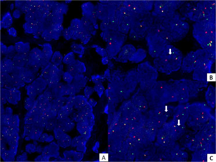
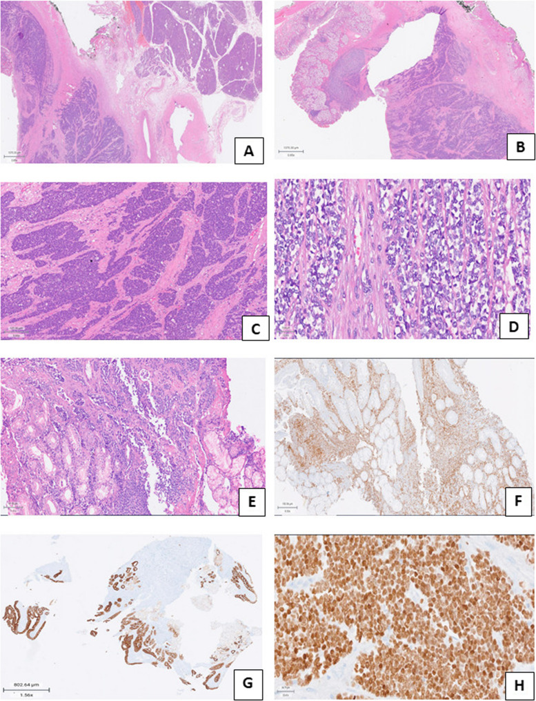
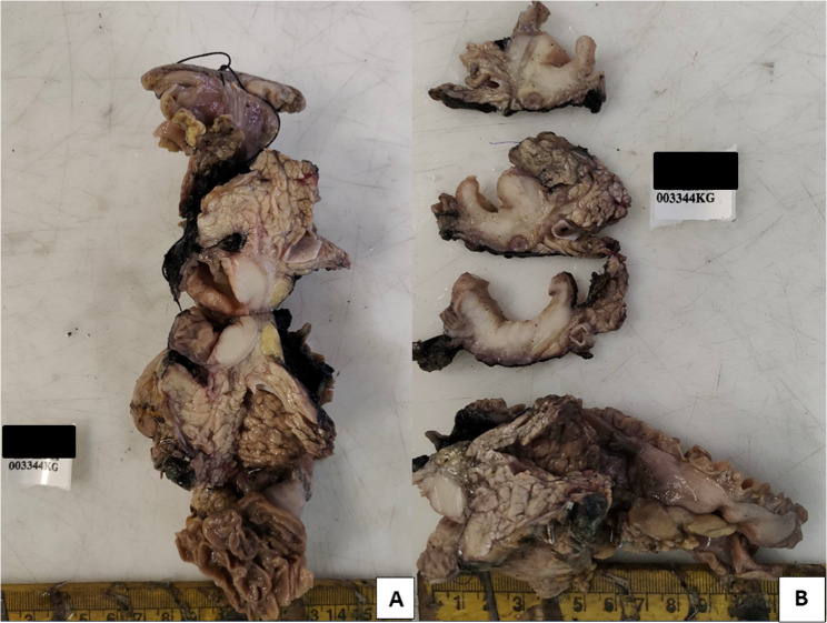
Ewing's sarcoma (ES) is exceptionally rare in the gastrointestinal tract, with only a few cases documented in the literature. We present a case of duodenal Ewing's sarcoma in a 44-year-old woman. The patient presented with severe abdominal pain, vomiting, and loss of appetite. Upper gastrointestinal endoscopy revealed a huge bulge in the first part of the duodenum (D1) wall, resulting in luminal narrowing in D1 and D2. A contrast-enhanced computed tomography scan corroborated the findings with no evidence of any distal metastasis. The patient is a known hypothyroid and had undergone hysterectomy five years back and cholecystectomy one year back. The patient underwent a Whipple procedure along with lymph node dissection. During surgery, a 4.5 cm mass was discovered in the D1 extending to the D2 segment of the duodenum. Histological examination showed a small round cell tumor in the submucosal region. The morphological differentials considered were gastrointestinal stromal tumor (GIST), poorly differentiated carcinoma, ES, neuroendocrine carcinoma (NEC) and lymphoma. Immunohistochemically the tumor cells tested positive for vimentin, NKX2.2, and CD99 (MIC2), and negative for markers of epithelial malignancy (AE1/AE3), lymphoma (CD45), melanoma (HMB45), NEC (INMS1,synaptophysin, chromogranin) and GIST (c-kit). One of the peri-duodenal lymph nodes showed a metastatic deposit. The case was diagnosed as Ewing's sarcoma (extra skeletal), TNM stage pT1 pN1. It was further ratified through fluorescence in situ hybridization (FISH), which showed a EWSR1 (22q12.1) gene rearrangement. A post-surgery PET scan indicated no residual disease. The patient had been receiving post-surgical adjuvant therapy and has completed four cycles of VAC (vincristine dactinomycin cyclophosphamide) and two cycles of VIME (Vincristine, Ifosfamide, Mesna, and Etoposide). The patient is disease-free with one-year follow-up. Follow up with PET scan is being performed at 3 months interval for the first year. The prognosis for Ewing's sarcoma in the gastrointestinal tract is not well-documented; however, a recent review of cases involving the small intestine suggests a poor prognosis. A follow up PET scanning every three months initially would help to target early recurrences and the interval can be increased after 2 years. Also emerging techniques like liquid biopsies is becoming increasingly relevant for detection of recurrences and metastasis.
期刊介绍:
World Journal of Surgical Oncology publishes articles related to surgical oncology and its allied subjects, such as epidemiology, cancer research, biomarkers, prevention, pathology, radiology, cancer treatment, clinical trials, multimodality treatment and molecular biology. Emphasis is placed on original research articles. The journal also publishes significant clinical case reports, as well as balanced and timely reviews on selected topics.
Oncology is a multidisciplinary super-speciality of which surgical oncology forms an integral component, especially with solid tumors. Surgical oncologists around the world are involved in research extending from detecting the mechanisms underlying the causation of cancer, to its treatment and prevention. The role of a surgical oncologist extends across the whole continuum of care. With continued developments in diagnosis and treatment, the role of a surgical oncologist is ever-changing. Hence, World Journal of Surgical Oncology aims to keep readers abreast with latest developments that will ultimately influence the work of surgical oncologists.

 求助内容:
求助内容: 应助结果提醒方式:


