Anna Jöbstl, Piera Maria Tierno, Anna-Katharina Gerstner, Gudrun Maria Feuchtner, Benedikt Schaefer, Herbert Tilg, Gerlig Widmann
{"title":"通过半自动化定量HCC病变增强LI-RADS。","authors":"Anna Jöbstl, Piera Maria Tierno, Anna-Katharina Gerstner, Gudrun Maria Feuchtner, Benedikt Schaefer, Herbert Tilg, Gerlig Widmann","doi":"10.3390/jpm15090400","DOIUrl":null,"url":null,"abstract":"<p><strong>Background/objectives: </strong>Hepatocellular carcinoma (HCC) is the most common primary malignant tumour of the liver. In a cirrhotic liver, each nodule larger than 10 mm demands further work-up using CT or MRI. The Liver Imaging Reporting and Data System (LI-RADS) is still based on visual assessment and measurements. The purpose of this study was to evaluate whether semi-automated quantification of visual LR-5 lesions is appropriate and can objectify HCC classification for personalized radiomic research.</p><p><strong>Methods: </strong>A total of 52 HCC patients (median age 67 years, 17% females, 83% males) from a retrospective data collection were evaluated visually and compared by the results using an oncology software with features of LI-RADS-based structured tumour evaluation and documentation, semi-automated tumour segmentation, and texture analysis.</p><p><strong>Results: </strong>Software-based evaluation of non-rim arterial-phase hyperenhancement (APHE) and non-peripheral washout, as well as the LI-RADS-score, showed no statistically significant differences compared with visual assessment (<i>p</i> = 0.2, 0.7, 0.17), with a consensus between a human reader and the software approach in 98% (APHE), 89% (washout), and 93% (threshold growth) of cases, respectively. The software provided automated LI-RADS classification, structured reporting, and quantitative features for HCC registries and radiomic research.</p><p><strong>Conclusions: </strong>The presented work may serve as an outlook for LI-RADS-based automated qualitative and quantitative evaluation. Future research may show if texture analysis can be used to foster personalized medical approaches in HCC.</p>","PeriodicalId":16722,"journal":{"name":"Journal of Personalized Medicine","volume":"15 9","pages":""},"PeriodicalIF":3.0000,"publicationDate":"2025-08-29","publicationTypes":"Journal Article","fieldsOfStudy":null,"isOpenAccess":false,"openAccessPdf":"https://www.ncbi.nlm.nih.gov/pmc/articles/PMC12470679/pdf/","citationCount":"0","resultStr":"{\"title\":\"Enhancing LI-RADS Through Semi-Automated Quantification of HCC Lesions.\",\"authors\":\"Anna Jöbstl, Piera Maria Tierno, Anna-Katharina Gerstner, Gudrun Maria Feuchtner, Benedikt Schaefer, Herbert Tilg, Gerlig Widmann\",\"doi\":\"10.3390/jpm15090400\",\"DOIUrl\":null,\"url\":null,\"abstract\":\"<p><strong>Background/objectives: </strong>Hepatocellular carcinoma (HCC) is the most common primary malignant tumour of the liver. In a cirrhotic liver, each nodule larger than 10 mm demands further work-up using CT or MRI. The Liver Imaging Reporting and Data System (LI-RADS) is still based on visual assessment and measurements. The purpose of this study was to evaluate whether semi-automated quantification of visual LR-5 lesions is appropriate and can objectify HCC classification for personalized radiomic research.</p><p><strong>Methods: </strong>A total of 52 HCC patients (median age 67 years, 17% females, 83% males) from a retrospective data collection were evaluated visually and compared by the results using an oncology software with features of LI-RADS-based structured tumour evaluation and documentation, semi-automated tumour segmentation, and texture analysis.</p><p><strong>Results: </strong>Software-based evaluation of non-rim arterial-phase hyperenhancement (APHE) and non-peripheral washout, as well as the LI-RADS-score, showed no statistically significant differences compared with visual assessment (<i>p</i> = 0.2, 0.7, 0.17), with a consensus between a human reader and the software approach in 98% (APHE), 89% (washout), and 93% (threshold growth) of cases, respectively. The software provided automated LI-RADS classification, structured reporting, and quantitative features for HCC registries and radiomic research.</p><p><strong>Conclusions: </strong>The presented work may serve as an outlook for LI-RADS-based automated qualitative and quantitative evaluation. Future research may show if texture analysis can be used to foster personalized medical approaches in HCC.</p>\",\"PeriodicalId\":16722,\"journal\":{\"name\":\"Journal of Personalized Medicine\",\"volume\":\"15 9\",\"pages\":\"\"},\"PeriodicalIF\":3.0000,\"publicationDate\":\"2025-08-29\",\"publicationTypes\":\"Journal Article\",\"fieldsOfStudy\":null,\"isOpenAccess\":false,\"openAccessPdf\":\"https://www.ncbi.nlm.nih.gov/pmc/articles/PMC12470679/pdf/\",\"citationCount\":\"0\",\"resultStr\":null,\"platform\":\"Semanticscholar\",\"paperid\":null,\"PeriodicalName\":\"Journal of Personalized Medicine\",\"FirstCategoryId\":\"3\",\"ListUrlMain\":\"https://doi.org/10.3390/jpm15090400\",\"RegionNum\":3,\"RegionCategory\":\"医学\",\"ArticlePicture\":[],\"TitleCN\":null,\"AbstractTextCN\":null,\"PMCID\":null,\"EPubDate\":\"\",\"PubModel\":\"\",\"JCR\":\"Q2\",\"JCRName\":\"HEALTH CARE SCIENCES & SERVICES\",\"Score\":null,\"Total\":0}","platform":"Semanticscholar","paperid":null,"PeriodicalName":"Journal of Personalized Medicine","FirstCategoryId":"3","ListUrlMain":"https://doi.org/10.3390/jpm15090400","RegionNum":3,"RegionCategory":"医学","ArticlePicture":[],"TitleCN":null,"AbstractTextCN":null,"PMCID":null,"EPubDate":"","PubModel":"","JCR":"Q2","JCRName":"HEALTH CARE SCIENCES & SERVICES","Score":null,"Total":0}
Enhancing LI-RADS Through Semi-Automated Quantification of HCC Lesions.
Background/objectives: Hepatocellular carcinoma (HCC) is the most common primary malignant tumour of the liver. In a cirrhotic liver, each nodule larger than 10 mm demands further work-up using CT or MRI. The Liver Imaging Reporting and Data System (LI-RADS) is still based on visual assessment and measurements. The purpose of this study was to evaluate whether semi-automated quantification of visual LR-5 lesions is appropriate and can objectify HCC classification for personalized radiomic research.
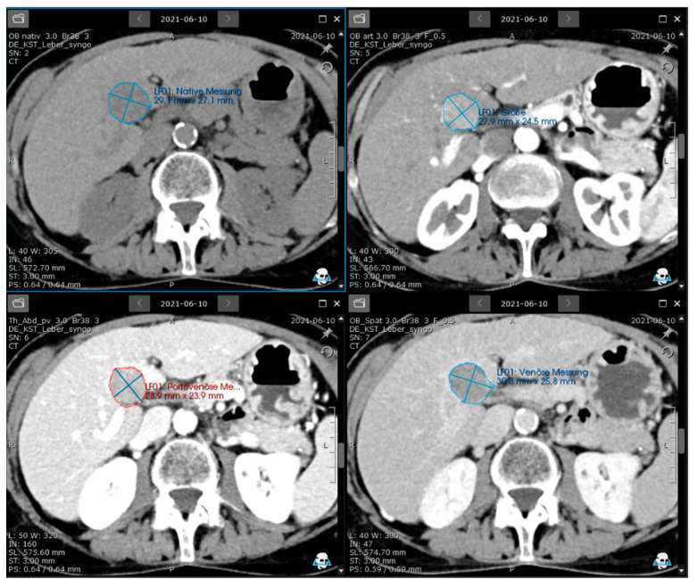
Methods: A total of 52 HCC patients (median age 67 years, 17% females, 83% males) from a retrospective data collection were evaluated visually and compared by the results using an oncology software with features of LI-RADS-based structured tumour evaluation and documentation, semi-automated tumour segmentation, and texture analysis.
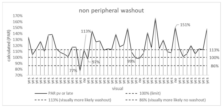
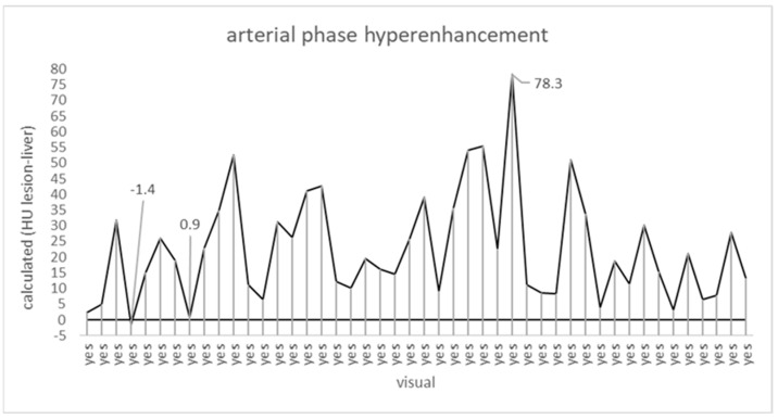
Results: Software-based evaluation of non-rim arterial-phase hyperenhancement (APHE) and non-peripheral washout, as well as the LI-RADS-score, showed no statistically significant differences compared with visual assessment (p = 0.2, 0.7, 0.17), with a consensus between a human reader and the software approach in 98% (APHE), 89% (washout), and 93% (threshold growth) of cases, respectively. The software provided automated LI-RADS classification, structured reporting, and quantitative features for HCC registries and radiomic research.
Conclusions: The presented work may serve as an outlook for LI-RADS-based automated qualitative and quantitative evaluation. Future research may show if texture analysis can be used to foster personalized medical approaches in HCC.
期刊介绍:
Journal of Personalized Medicine (JPM; ISSN 2075-4426) is an international, open access journal aimed at bringing all aspects of personalized medicine to one platform. JPM publishes cutting edge, innovative preclinical and translational scientific research and technologies related to personalized medicine (e.g., pharmacogenomics/proteomics, systems biology). JPM recognizes that personalized medicine—the assessment of genetic, environmental and host factors that cause variability of individuals—is a challenging, transdisciplinary topic that requires discussions from a range of experts. For a comprehensive perspective of personalized medicine, JPM aims to integrate expertise from the molecular and translational sciences, therapeutics and diagnostics, as well as discussions of regulatory, social, ethical and policy aspects. We provide a forum to bring together academic and clinical researchers, biotechnology, diagnostic and pharmaceutical companies, health professionals, regulatory and ethical experts, and government and regulatory authorities.
 求助内容:
求助内容: 应助结果提醒方式:


