Prajitha Biju, Shaila Lewis, Manjunath M. Shenoy, Ashwini Prabhu, Ranajit Das, Anne Boyina Sravani, Mohammed Gulzar Ahmed, Vivek Ghate
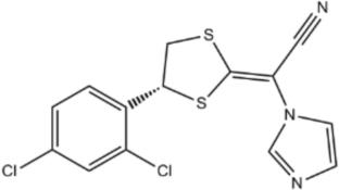
{"title":"有效和可持续的靶向给药Luliconazole治疗足癣感染的Niosomes","authors":"Prajitha Biju, Shaila Lewis, Manjunath M. Shenoy, Ashwini Prabhu, Ranajit Das, Anne Boyina Sravani, Mohammed Gulzar Ahmed, Vivek Ghate","doi":"10.1007/s10876-025-02853-8","DOIUrl":null,"url":null,"abstract":"<div><p>A simple and cost-effective method—the ‘Vial Method’ was utilized to develop niosomes incorporating luliconazole, offering a convenient alternative to the traditional thin-layer hydration method. Niosomes were prepared using the ‘Vial Method’ with nonionic surfactants and cholesterol. The method was optimized using the Design-Expert® software to refine particle size and the entrapment efficiency, with the morphology of niosomes analyzed through Transmission Electron Microscopy. The compatibility and stability of the components and their physical mixture were assessed using infrared spectroscopy and Differential Scanning Calorimetry. Antifungal activity was determined by the disc diffusion method, with <i>in vitro</i> drug release studies, and cytocompatibility testing, and acute skin irritation tests performed <i>in vivo</i>. The optimized niosomes were spherical, with a size of ~221.0 nm, drug entrapment efficiency of ~94%, and drug loading capacity of approximately 28%. In comparison, the conventional method produced vesicles of ~275.0 nm size and a drug entrapment efficiency of ~97%. The niosomes developed using the simple method showed an initial burst release followed by sustained release, with release data fitting best with zero order kinetics. The optimized niosomes demonstrated significant antifungal potential against <i>Candida</i> strains (p < 0.05), and <i>in vivo</i> skin irritation tests confirmed their safety. Luliconzole niosomes were efficiently prepared using the ‘Vial Method’ and demonstrated for its non-irritancy, and possibility of effective management of tinea infections caused by <i>Candida</i> species.</p><h3>Graphical Abstract</h3>\n<div><figure><div><div><picture><source><img></source></picture></div></div></figure></div></div>","PeriodicalId":618,"journal":{"name":"Journal of Cluster Science","volume":"36 4","pages":""},"PeriodicalIF":3.6000,"publicationDate":"2025-06-10","publicationTypes":"Journal Article","fieldsOfStudy":null,"isOpenAccess":false,"openAccessPdf":"","citationCount":"0","resultStr":"{\"title\":\"Efficient and Sustainable Niosomes for Targeted Delivery of Luliconazole in the Treatment of Tinea Infections\",\"authors\":\"Prajitha Biju, Shaila Lewis, Manjunath M. Shenoy, Ashwini Prabhu, Ranajit Das, Anne Boyina Sravani, Mohammed Gulzar Ahmed, Vivek Ghate\",\"doi\":\"10.1007/s10876-025-02853-8\",\"DOIUrl\":null,\"url\":null,\"abstract\":\"<div><p>A simple and cost-effective method—the ‘Vial Method’ was utilized to develop niosomes incorporating luliconazole, offering a convenient alternative to the traditional thin-layer hydration method. Niosomes were prepared using the ‘Vial Method’ with nonionic surfactants and cholesterol. The method was optimized using the Design-Expert® software to refine particle size and the entrapment efficiency, with the morphology of niosomes analyzed through Transmission Electron Microscopy. The compatibility and stability of the components and their physical mixture were assessed using infrared spectroscopy and Differential Scanning Calorimetry. Antifungal activity was determined by the disc diffusion method, with <i>in vitro</i> drug release studies, and cytocompatibility testing, and acute skin irritation tests performed <i>in vivo</i>. The optimized niosomes were spherical, with a size of ~221.0 nm, drug entrapment efficiency of ~94%, and drug loading capacity of approximately 28%. In comparison, the conventional method produced vesicles of ~275.0 nm size and a drug entrapment efficiency of ~97%. The niosomes developed using the simple method showed an initial burst release followed by sustained release, with release data fitting best with zero order kinetics. The optimized niosomes demonstrated significant antifungal potential against <i>Candida</i> strains (p < 0.05), and <i>in vivo</i> skin irritation tests confirmed their safety. Luliconzole niosomes were efficiently prepared using the ‘Vial Method’ and demonstrated for its non-irritancy, and possibility of effective management of tinea infections caused by <i>Candida</i> species.</p><h3>Graphical Abstract</h3>\\n<div><figure><div><div><picture><source><img></source></picture></div></div></figure></div></div>\",\"PeriodicalId\":618,\"journal\":{\"name\":\"Journal of Cluster Science\",\"volume\":\"36 4\",\"pages\":\"\"},\"PeriodicalIF\":3.6000,\"publicationDate\":\"2025-06-10\",\"publicationTypes\":\"Journal Article\",\"fieldsOfStudy\":null,\"isOpenAccess\":false,\"openAccessPdf\":\"\",\"citationCount\":\"0\",\"resultStr\":null,\"platform\":\"Semanticscholar\",\"paperid\":null,\"PeriodicalName\":\"Journal of Cluster Science\",\"FirstCategoryId\":\"92\",\"ListUrlMain\":\"https://link.springer.com/article/10.1007/s10876-025-02853-8\",\"RegionNum\":4,\"RegionCategory\":\"化学\",\"ArticlePicture\":[],\"TitleCN\":null,\"AbstractTextCN\":null,\"PMCID\":null,\"EPubDate\":\"\",\"PubModel\":\"\",\"JCR\":\"Q2\",\"JCRName\":\"CHEMISTRY, INORGANIC & NUCLEAR\",\"Score\":null,\"Total\":0}","platform":"Semanticscholar","paperid":null,"PeriodicalName":"Journal of Cluster Science","FirstCategoryId":"92","ListUrlMain":"https://link.springer.com/article/10.1007/s10876-025-02853-8","RegionNum":4,"RegionCategory":"化学","ArticlePicture":[],"TitleCN":null,"AbstractTextCN":null,"PMCID":null,"EPubDate":"","PubModel":"","JCR":"Q2","JCRName":"CHEMISTRY, INORGANIC & NUCLEAR","Score":null,"Total":0}
Efficient and Sustainable Niosomes for Targeted Delivery of Luliconazole in the Treatment of Tinea Infections
A simple and cost-effective method—the ‘Vial Method’ was utilized to develop niosomes incorporating luliconazole, offering a convenient alternative to the traditional thin-layer hydration method. Niosomes were prepared using the ‘Vial Method’ with nonionic surfactants and cholesterol. The method was optimized using the Design-Expert® software to refine particle size and the entrapment efficiency, with the morphology of niosomes analyzed through Transmission Electron Microscopy. The compatibility and stability of the components and their physical mixture were assessed using infrared spectroscopy and Differential Scanning Calorimetry. Antifungal activity was determined by the disc diffusion method, with in vitro drug release studies, and cytocompatibility testing, and acute skin irritation tests performed in vivo. The optimized niosomes were spherical, with a size of ~221.0 nm, drug entrapment efficiency of ~94%, and drug loading capacity of approximately 28%. In comparison, the conventional method produced vesicles of ~275.0 nm size and a drug entrapment efficiency of ~97%. The niosomes developed using the simple method showed an initial burst release followed by sustained release, with release data fitting best with zero order kinetics. The optimized niosomes demonstrated significant antifungal potential against Candida strains (p < 0.05), and in vivo skin irritation tests confirmed their safety. Luliconzole niosomes were efficiently prepared using the ‘Vial Method’ and demonstrated for its non-irritancy, and possibility of effective management of tinea infections caused by Candida species.
期刊介绍:
The journal publishes the following types of papers: (a) original and important research;
(b) authoritative comprehensive reviews or short overviews of topics of current
interest; (c) brief but urgent communications on new significant research; and (d)
commentaries intended to foster the exchange of innovative or provocative ideas, and
to encourage dialogue, amongst researchers working in different cluster
disciplines.
 求助内容:
求助内容: 应助结果提醒方式:


