Abdulaziz AlTaweel, Faisal Joueidi, Ahmad Joueidi, Ahmed AlDhubaiki, Hamad Mohammed Qabha, Homoud Abdulaziz AlZaid
{"title":"对比增强超声在早期肝细胞癌诊断中的有效性评价:系统综述。","authors":"Abdulaziz AlTaweel, Faisal Joueidi, Ahmad Joueidi, Ahmed AlDhubaiki, Hamad Mohammed Qabha, Homoud Abdulaziz AlZaid","doi":"10.3389/fradi.2025.1661522","DOIUrl":null,"url":null,"abstract":"<p><strong>Objectives: </strong>To investigate the evaluation of the effectiveness of contrast-enhanced ultrasound (CEUS) in the diagnosis of small hepatocellular carcinoma (HCC).</p><p><strong>Methods: </strong>A thorough search was conducted for pertinent literature using PubMed, SCOPUS, Web of Science, Science Direct, and Wiley Library. Rayyan QRCI was used throughout this extensive procedure.</p><p><strong>Results: </strong>Our results included thirteen studies with a total of 2016 patients, and 1672 (82.9%) were males. The follow-up duration ranged from 3 months to 24 months. CEUS was useful in anticipating the early recurrence of HCC, predicting the early recurrence of solitary lesion HCC patients, and differentiating between HCC and intrahepatic cholangiocarcinoma <3 Cm, distinguishing HCC from dysplastic nodules from tiny liver nodules, CEUS in cirrhotic patients. When paired with CEUS, conventional ultrasonography can detect minor HCC and assist in patient monitoring for those who receive an early diagnosis of HCC. CEUS showed high concordance with CECT for diagnosing lesions 2.1-3.0 cm in size. Notable limitations included heterogeneity in protocols and predominance of Asian populations (12/13 studies).</p><p><strong>Conclusion: </strong>CEUS offers significant clinical value as a noninvasive diagnostic tool, particularly for 1-3 cm lesions in cirrhotic patients and cases where CT is contraindicated, though protocol standardization and Western population validation remain needed.</p>","PeriodicalId":73101,"journal":{"name":"Frontiers in radiology","volume":"5 ","pages":"1661522"},"PeriodicalIF":2.3000,"publicationDate":"2025-09-09","publicationTypes":"Journal Article","fieldsOfStudy":null,"isOpenAccess":false,"openAccessPdf":"https://www.ncbi.nlm.nih.gov/pmc/articles/PMC12454341/pdf/","citationCount":"0","resultStr":"{\"title\":\"Evaluation of the effectiveness of contrast-enhanced ultrasound in the diagnosis of early hepatocellular carcinoma: a systematic review.\",\"authors\":\"Abdulaziz AlTaweel, Faisal Joueidi, Ahmad Joueidi, Ahmed AlDhubaiki, Hamad Mohammed Qabha, Homoud Abdulaziz AlZaid\",\"doi\":\"10.3389/fradi.2025.1661522\",\"DOIUrl\":null,\"url\":null,\"abstract\":\"<p><strong>Objectives: </strong>To investigate the evaluation of the effectiveness of contrast-enhanced ultrasound (CEUS) in the diagnosis of small hepatocellular carcinoma (HCC).</p><p><strong>Methods: </strong>A thorough search was conducted for pertinent literature using PubMed, SCOPUS, Web of Science, Science Direct, and Wiley Library. Rayyan QRCI was used throughout this extensive procedure.</p><p><strong>Results: </strong>Our results included thirteen studies with a total of 2016 patients, and 1672 (82.9%) were males. The follow-up duration ranged from 3 months to 24 months. CEUS was useful in anticipating the early recurrence of HCC, predicting the early recurrence of solitary lesion HCC patients, and differentiating between HCC and intrahepatic cholangiocarcinoma <3 Cm, distinguishing HCC from dysplastic nodules from tiny liver nodules, CEUS in cirrhotic patients. When paired with CEUS, conventional ultrasonography can detect minor HCC and assist in patient monitoring for those who receive an early diagnosis of HCC. CEUS showed high concordance with CECT for diagnosing lesions 2.1-3.0 cm in size. Notable limitations included heterogeneity in protocols and predominance of Asian populations (12/13 studies).</p><p><strong>Conclusion: </strong>CEUS offers significant clinical value as a noninvasive diagnostic tool, particularly for 1-3 cm lesions in cirrhotic patients and cases where CT is contraindicated, though protocol standardization and Western population validation remain needed.</p>\",\"PeriodicalId\":73101,\"journal\":{\"name\":\"Frontiers in radiology\",\"volume\":\"5 \",\"pages\":\"1661522\"},\"PeriodicalIF\":2.3000,\"publicationDate\":\"2025-09-09\",\"publicationTypes\":\"Journal Article\",\"fieldsOfStudy\":null,\"isOpenAccess\":false,\"openAccessPdf\":\"https://www.ncbi.nlm.nih.gov/pmc/articles/PMC12454341/pdf/\",\"citationCount\":\"0\",\"resultStr\":null,\"platform\":\"Semanticscholar\",\"paperid\":null,\"PeriodicalName\":\"Frontiers in radiology\",\"FirstCategoryId\":\"1085\",\"ListUrlMain\":\"https://doi.org/10.3389/fradi.2025.1661522\",\"RegionNum\":0,\"RegionCategory\":null,\"ArticlePicture\":[],\"TitleCN\":null,\"AbstractTextCN\":null,\"PMCID\":null,\"EPubDate\":\"2025/1/1 0:00:00\",\"PubModel\":\"eCollection\",\"JCR\":\"\",\"JCRName\":\"\",\"Score\":null,\"Total\":0}","platform":"Semanticscholar","paperid":null,"PeriodicalName":"Frontiers in radiology","FirstCategoryId":"1085","ListUrlMain":"https://doi.org/10.3389/fradi.2025.1661522","RegionNum":0,"RegionCategory":null,"ArticlePicture":[],"TitleCN":null,"AbstractTextCN":null,"PMCID":null,"EPubDate":"2025/1/1 0:00:00","PubModel":"eCollection","JCR":"","JCRName":"","Score":null,"Total":0}
引用次数: 0
摘要
目的:探讨超声造影(CEUS)在小肝癌(HCC)诊断中的价值。方法:检索PubMed、SCOPUS、Web of Science、Science Direct、Wiley Library等相关文献。Rayyan QRCI在整个广泛的程序中使用。结果:纳入13项研究,共纳入2016例患者,其中男性1672例(82.9%)。随访时间3 ~ 24个月。结论:超声造影作为一种无创诊断工具具有重要的临床价值,特别是对于肝硬化患者1-3 cm病变和CT禁忌的病例,尽管仍需要方案标准化和西方人群验证。
Evaluation of the effectiveness of contrast-enhanced ultrasound in the diagnosis of early hepatocellular carcinoma: a systematic review.
Objectives: To investigate the evaluation of the effectiveness of contrast-enhanced ultrasound (CEUS) in the diagnosis of small hepatocellular carcinoma (HCC).
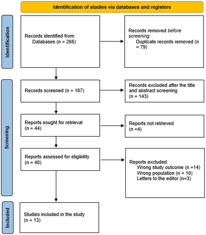
Methods: A thorough search was conducted for pertinent literature using PubMed, SCOPUS, Web of Science, Science Direct, and Wiley Library. Rayyan QRCI was used throughout this extensive procedure.
Results: Our results included thirteen studies with a total of 2016 patients, and 1672 (82.9%) were males. The follow-up duration ranged from 3 months to 24 months. CEUS was useful in anticipating the early recurrence of HCC, predicting the early recurrence of solitary lesion HCC patients, and differentiating between HCC and intrahepatic cholangiocarcinoma <3 Cm, distinguishing HCC from dysplastic nodules from tiny liver nodules, CEUS in cirrhotic patients. When paired with CEUS, conventional ultrasonography can detect minor HCC and assist in patient monitoring for those who receive an early diagnosis of HCC. CEUS showed high concordance with CECT for diagnosing lesions 2.1-3.0 cm in size. Notable limitations included heterogeneity in protocols and predominance of Asian populations (12/13 studies).
Conclusion: CEUS offers significant clinical value as a noninvasive diagnostic tool, particularly for 1-3 cm lesions in cirrhotic patients and cases where CT is contraindicated, though protocol standardization and Western population validation remain needed.

 求助内容:
求助内容: 应助结果提醒方式:


