Yuan-Kai Fu, Matthew Lin, Kuo-Hsuan Hung, Lung-Kun Yeh, Hsin-Yuan Tan
{"title":"内皮损伤后角膜内皮单细胞大量丢失。","authors":"Yuan-Kai Fu, Matthew Lin, Kuo-Hsuan Hung, Lung-Kun Yeh, Hsin-Yuan Tan","doi":"10.4103/tjo.TJO-D-24-00118","DOIUrl":null,"url":null,"abstract":"<p><strong>Purpose: </strong>The purpose of this study was to investigate corneal endothelial changes following common clinical endothelial injury scenarios in order to uncover mechanisms underlying unexplained chronic corneal endothelial wound healing.</p><p><strong>Materials and methods: </strong>This cross-sectional study included patients with endothelial injuries from three common scenarios: postcataract surgery, corneal dystrophies, and penetrating injuries. Noncontact specular microscopy was used to capture images from five distinct corneal regions. Endothelial cell density (ECD), coefficient of variation (CV), and percentage of hexagonal cells (HEX) were assessed. All endothelial photographs were also reviewed. Statistical analysis was performed to compare injured and noninjured eyes.</p><p><strong>Results: </strong>Seventy-seven patients were enrolled, with a mean age of 64 years (48 females, 29 males). The mean central ECD was 2138.91 ± 869.34 cells/mm<sup>2</sup> in postcataract surgery eyes, 1999.48 ± 763.91 cells/mm<sup>2</sup> in endothelial dystrophy eyes, and 1854.86 ± 551.85 cells/mm<sup>2</sup> in trauma cases. While most parameters showed no significant differences, postcataract surgery eyes exhibited a significant increase in CV value in the upper and temporal regions (<i>P</i> < 0.05). Unexpectedly, stochastic single-cell loss was observed in 42.86% of patients, continuing up to two years postinjury. This loss was significantly higher compared to uninjured eyes (<i>P</i> = 0.00005), suggesting that excessive single-cell loss occurs well beyond the expected wound healing period.</p><p><strong>Conclusion: </strong>We identified accelerated stochastic single-cell loss in the corneal endothelium following primary injuries, persisting well beyond the expected wound healing period, a phenomenon that has not been previously highlighted. This finding offers a potential explanation for the chronic endothelial cell loss following a primary injury.</p>","PeriodicalId":44978,"journal":{"name":"Taiwan Journal of Ophthalmology","volume":"15 3","pages":"480-486"},"PeriodicalIF":1.2000,"publicationDate":"2025-05-30","publicationTypes":"Journal Article","fieldsOfStudy":null,"isOpenAccess":false,"openAccessPdf":"https://www.ncbi.nlm.nih.gov/pmc/articles/PMC12456922/pdf/","citationCount":"0","resultStr":"{\"title\":\"Excessive corneal endothelial single-cell loss following endothelial injuries.\",\"authors\":\"Yuan-Kai Fu, Matthew Lin, Kuo-Hsuan Hung, Lung-Kun Yeh, Hsin-Yuan Tan\",\"doi\":\"10.4103/tjo.TJO-D-24-00118\",\"DOIUrl\":null,\"url\":null,\"abstract\":\"<p><strong>Purpose: </strong>The purpose of this study was to investigate corneal endothelial changes following common clinical endothelial injury scenarios in order to uncover mechanisms underlying unexplained chronic corneal endothelial wound healing.</p><p><strong>Materials and methods: </strong>This cross-sectional study included patients with endothelial injuries from three common scenarios: postcataract surgery, corneal dystrophies, and penetrating injuries. Noncontact specular microscopy was used to capture images from five distinct corneal regions. Endothelial cell density (ECD), coefficient of variation (CV), and percentage of hexagonal cells (HEX) were assessed. All endothelial photographs were also reviewed. Statistical analysis was performed to compare injured and noninjured eyes.</p><p><strong>Results: </strong>Seventy-seven patients were enrolled, with a mean age of 64 years (48 females, 29 males). The mean central ECD was 2138.91 ± 869.34 cells/mm<sup>2</sup> in postcataract surgery eyes, 1999.48 ± 763.91 cells/mm<sup>2</sup> in endothelial dystrophy eyes, and 1854.86 ± 551.85 cells/mm<sup>2</sup> in trauma cases. While most parameters showed no significant differences, postcataract surgery eyes exhibited a significant increase in CV value in the upper and temporal regions (<i>P</i> < 0.05). Unexpectedly, stochastic single-cell loss was observed in 42.86% of patients, continuing up to two years postinjury. This loss was significantly higher compared to uninjured eyes (<i>P</i> = 0.00005), suggesting that excessive single-cell loss occurs well beyond the expected wound healing period.</p><p><strong>Conclusion: </strong>We identified accelerated stochastic single-cell loss in the corneal endothelium following primary injuries, persisting well beyond the expected wound healing period, a phenomenon that has not been previously highlighted. This finding offers a potential explanation for the chronic endothelial cell loss following a primary injury.</p>\",\"PeriodicalId\":44978,\"journal\":{\"name\":\"Taiwan Journal of Ophthalmology\",\"volume\":\"15 3\",\"pages\":\"480-486\"},\"PeriodicalIF\":1.2000,\"publicationDate\":\"2025-05-30\",\"publicationTypes\":\"Journal Article\",\"fieldsOfStudy\":null,\"isOpenAccess\":false,\"openAccessPdf\":\"https://www.ncbi.nlm.nih.gov/pmc/articles/PMC12456922/pdf/\",\"citationCount\":\"0\",\"resultStr\":null,\"platform\":\"Semanticscholar\",\"paperid\":null,\"PeriodicalName\":\"Taiwan Journal of Ophthalmology\",\"FirstCategoryId\":\"1085\",\"ListUrlMain\":\"https://doi.org/10.4103/tjo.TJO-D-24-00118\",\"RegionNum\":0,\"RegionCategory\":null,\"ArticlePicture\":[],\"TitleCN\":null,\"AbstractTextCN\":null,\"PMCID\":null,\"EPubDate\":\"2025/7/1 0:00:00\",\"PubModel\":\"eCollection\",\"JCR\":\"Q4\",\"JCRName\":\"OPHTHALMOLOGY\",\"Score\":null,\"Total\":0}","platform":"Semanticscholar","paperid":null,"PeriodicalName":"Taiwan Journal of Ophthalmology","FirstCategoryId":"1085","ListUrlMain":"https://doi.org/10.4103/tjo.TJO-D-24-00118","RegionNum":0,"RegionCategory":null,"ArticlePicture":[],"TitleCN":null,"AbstractTextCN":null,"PMCID":null,"EPubDate":"2025/7/1 0:00:00","PubModel":"eCollection","JCR":"Q4","JCRName":"OPHTHALMOLOGY","Score":null,"Total":0}
Excessive corneal endothelial single-cell loss following endothelial injuries.
Purpose: The purpose of this study was to investigate corneal endothelial changes following common clinical endothelial injury scenarios in order to uncover mechanisms underlying unexplained chronic corneal endothelial wound healing.
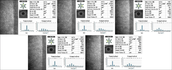
Materials and methods: This cross-sectional study included patients with endothelial injuries from three common scenarios: postcataract surgery, corneal dystrophies, and penetrating injuries. Noncontact specular microscopy was used to capture images from five distinct corneal regions. Endothelial cell density (ECD), coefficient of variation (CV), and percentage of hexagonal cells (HEX) were assessed. All endothelial photographs were also reviewed. Statistical analysis was performed to compare injured and noninjured eyes.
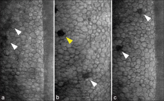
Results: Seventy-seven patients were enrolled, with a mean age of 64 years (48 females, 29 males). The mean central ECD was 2138.91 ± 869.34 cells/mm2 in postcataract surgery eyes, 1999.48 ± 763.91 cells/mm2 in endothelial dystrophy eyes, and 1854.86 ± 551.85 cells/mm2 in trauma cases. While most parameters showed no significant differences, postcataract surgery eyes exhibited a significant increase in CV value in the upper and temporal regions (P < 0.05). Unexpectedly, stochastic single-cell loss was observed in 42.86% of patients, continuing up to two years postinjury. This loss was significantly higher compared to uninjured eyes (P = 0.00005), suggesting that excessive single-cell loss occurs well beyond the expected wound healing period.
Conclusion: We identified accelerated stochastic single-cell loss in the corneal endothelium following primary injuries, persisting well beyond the expected wound healing period, a phenomenon that has not been previously highlighted. This finding offers a potential explanation for the chronic endothelial cell loss following a primary injury.

 求助内容:
求助内容: 应助结果提醒方式:


