{"title":"中央凹无血管区及周围毛细血管网的改变是遗传性黄斑营养不良视觉预后的重要指标。","authors":"Yen-Ching Lin, Ting-Chieh Ko, Chang-Hao Yang, Pei-Hsuan Chen, Chung-May Yang, Pei-Lung Chen, Bo-I Kuo, Ta-Ching Chen","doi":"10.4103/tjo.TJO-D-25-00089","DOIUrl":null,"url":null,"abstract":"<p><strong>Purpose: </strong>Hereditary macular dystrophy (MD) usually severely affects the central vision. This study aimed to explore macular microcirculation and its relationship with disease progression in different morphological patterns of MD.</p><p><strong>Materials and methods: </strong>Sixty-five patients with MD and 26 healthy participants were included. Panel-based next-generation sequencing (NGS), fundus autofluorescence (FAF), and optical coherence tomography angiography (OCTA) were used for genetic diagnosis, morphological classification, and evaluation of macular microcirculation, respectively. Patients were divided into two groups: the central lesion group (CLG) and the dispersed lesion group (DLG), based on FAF findings. The alterations in microcirculation between the groups and subgroups were analyzed and correlated with visual preservation.</p><p><strong>Results: </strong>A high diagnostic rate of disease-causing genes was achieved with a panel-based NGS test (72.3%). Compromised macular microcirculation was seen in MD of all genotypes. Enlargement of the foveal avascular zone and decreased foveal vessel density was significantly correlated with impaired vision (both <i>P</i> < 0.05). In Stargardt disease, the CLG had an earlier onset than the DLG, with more severely impaired central vision and compromised microcirculation.</p><p><strong>Conclusion: </strong>OCTA is a reliable, noninvasive tool for evaluating the microcirculation of MD. Our results demonstrate that compromised macular microcirculation occurs with MD, and foveal microcirculation is crucial for visual preservation.</p>","PeriodicalId":44978,"journal":{"name":"Taiwan Journal of Ophthalmology","volume":"15 3","pages":"457-465"},"PeriodicalIF":1.2000,"publicationDate":"2025-09-05","publicationTypes":"Journal Article","fieldsOfStudy":null,"isOpenAccess":false,"openAccessPdf":"https://www.ncbi.nlm.nih.gov/pmc/articles/PMC12456923/pdf/","citationCount":"0","resultStr":"{\"title\":\"Alterations in the foveal avascular zone and surrounding capillary network as important indicators of visual prognosis for hereditary macular dystrophy.\",\"authors\":\"Yen-Ching Lin, Ting-Chieh Ko, Chang-Hao Yang, Pei-Hsuan Chen, Chung-May Yang, Pei-Lung Chen, Bo-I Kuo, Ta-Ching Chen\",\"doi\":\"10.4103/tjo.TJO-D-25-00089\",\"DOIUrl\":null,\"url\":null,\"abstract\":\"<p><strong>Purpose: </strong>Hereditary macular dystrophy (MD) usually severely affects the central vision. This study aimed to explore macular microcirculation and its relationship with disease progression in different morphological patterns of MD.</p><p><strong>Materials and methods: </strong>Sixty-five patients with MD and 26 healthy participants were included. Panel-based next-generation sequencing (NGS), fundus autofluorescence (FAF), and optical coherence tomography angiography (OCTA) were used for genetic diagnosis, morphological classification, and evaluation of macular microcirculation, respectively. Patients were divided into two groups: the central lesion group (CLG) and the dispersed lesion group (DLG), based on FAF findings. The alterations in microcirculation between the groups and subgroups were analyzed and correlated with visual preservation.</p><p><strong>Results: </strong>A high diagnostic rate of disease-causing genes was achieved with a panel-based NGS test (72.3%). Compromised macular microcirculation was seen in MD of all genotypes. Enlargement of the foveal avascular zone and decreased foveal vessel density was significantly correlated with impaired vision (both <i>P</i> < 0.05). In Stargardt disease, the CLG had an earlier onset than the DLG, with more severely impaired central vision and compromised microcirculation.</p><p><strong>Conclusion: </strong>OCTA is a reliable, noninvasive tool for evaluating the microcirculation of MD. Our results demonstrate that compromised macular microcirculation occurs with MD, and foveal microcirculation is crucial for visual preservation.</p>\",\"PeriodicalId\":44978,\"journal\":{\"name\":\"Taiwan Journal of Ophthalmology\",\"volume\":\"15 3\",\"pages\":\"457-465\"},\"PeriodicalIF\":1.2000,\"publicationDate\":\"2025-09-05\",\"publicationTypes\":\"Journal Article\",\"fieldsOfStudy\":null,\"isOpenAccess\":false,\"openAccessPdf\":\"https://www.ncbi.nlm.nih.gov/pmc/articles/PMC12456923/pdf/\",\"citationCount\":\"0\",\"resultStr\":null,\"platform\":\"Semanticscholar\",\"paperid\":null,\"PeriodicalName\":\"Taiwan Journal of Ophthalmology\",\"FirstCategoryId\":\"1085\",\"ListUrlMain\":\"https://doi.org/10.4103/tjo.TJO-D-25-00089\",\"RegionNum\":0,\"RegionCategory\":null,\"ArticlePicture\":[],\"TitleCN\":null,\"AbstractTextCN\":null,\"PMCID\":null,\"EPubDate\":\"2025/7/1 0:00:00\",\"PubModel\":\"eCollection\",\"JCR\":\"Q4\",\"JCRName\":\"OPHTHALMOLOGY\",\"Score\":null,\"Total\":0}","platform":"Semanticscholar","paperid":null,"PeriodicalName":"Taiwan Journal of Ophthalmology","FirstCategoryId":"1085","ListUrlMain":"https://doi.org/10.4103/tjo.TJO-D-25-00089","RegionNum":0,"RegionCategory":null,"ArticlePicture":[],"TitleCN":null,"AbstractTextCN":null,"PMCID":null,"EPubDate":"2025/7/1 0:00:00","PubModel":"eCollection","JCR":"Q4","JCRName":"OPHTHALMOLOGY","Score":null,"Total":0}
Alterations in the foveal avascular zone and surrounding capillary network as important indicators of visual prognosis for hereditary macular dystrophy.
Purpose: Hereditary macular dystrophy (MD) usually severely affects the central vision. This study aimed to explore macular microcirculation and its relationship with disease progression in different morphological patterns of MD.
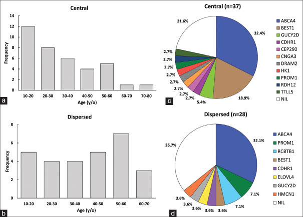
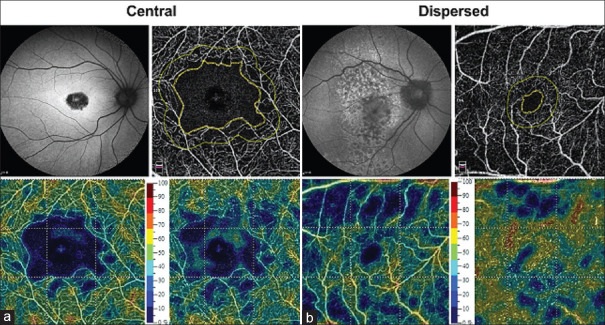
Materials and methods: Sixty-five patients with MD and 26 healthy participants were included. Panel-based next-generation sequencing (NGS), fundus autofluorescence (FAF), and optical coherence tomography angiography (OCTA) were used for genetic diagnosis, morphological classification, and evaluation of macular microcirculation, respectively. Patients were divided into two groups: the central lesion group (CLG) and the dispersed lesion group (DLG), based on FAF findings. The alterations in microcirculation between the groups and subgroups were analyzed and correlated with visual preservation.
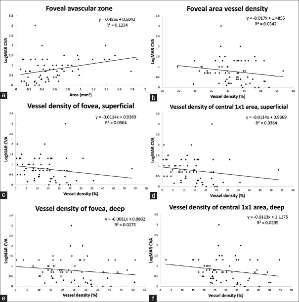
Results: A high diagnostic rate of disease-causing genes was achieved with a panel-based NGS test (72.3%). Compromised macular microcirculation was seen in MD of all genotypes. Enlargement of the foveal avascular zone and decreased foveal vessel density was significantly correlated with impaired vision (both P < 0.05). In Stargardt disease, the CLG had an earlier onset than the DLG, with more severely impaired central vision and compromised microcirculation.
Conclusion: OCTA is a reliable, noninvasive tool for evaluating the microcirculation of MD. Our results demonstrate that compromised macular microcirculation occurs with MD, and foveal microcirculation is crucial for visual preservation.

 求助内容:
求助内容: 应助结果提醒方式:


