Sabine Guehery, Julian Plenard, François Lafourcade, Franck Lapegue, Laurent Zabraniecki, Arnaud Constantin, Nicolas Sans, Adeline Ruyssen Witrand, Marie Faruch Bilfeld
{"title":"无脊柱关节炎的骶髂关节骨髓水肿:34例髂凝性骨炎的回顾性研究。","authors":"Sabine Guehery, Julian Plenard, François Lafourcade, Franck Lapegue, Laurent Zabraniecki, Arnaud Constantin, Nicolas Sans, Adeline Ruyssen Witrand, Marie Faruch Bilfeld","doi":"10.1093/rap/rkaf108","DOIUrl":null,"url":null,"abstract":"<p><strong>Objective: </strong>To determine the prevalence of bone marrow oedema (BME) in osteitis condensans ilii (OCI) on MRI of the sacroiliac joint (SIJ). Secondary objectives include comparisons of socio-demographic characteristics, prevalence of other imaging features (MRI, CT scan), and low back pain in patients with OCI with those in a sex- and age-matched control group.</p><p><strong>Methods: </strong>A total of 34 patients with OCI, including 29 with MRI, were recruited for retrospective analysis. The SIJ MRIs were retrospectively analysed by two readers. A sex- and age-matched control group of patients without SIJ disorders was included. In both groups, the presence of structural bone abnormalities was assessed by CT scan analysis, and socio-demographic data were obtained by telephone questionnaire. A longitudinal analysis was conducted on patients who had undergone multiple imaging examinations.</p><p><strong>Results: </strong>All patients were female with a mean age of 34 years. BME was observed in 66% (19/29) of OCI patients. BME in OCI was mainly located in the anterior-middle quadrant (43.48%). OCI patients had significantly more than one delivery (<i>P</i> = 0.0094, McNemar test), even if OCI was found in four nulliparous patients (15%). OCI patients experienced significantly more pain (<i>P</i> = 0.0026, McNemar test).</p><p><strong>Conclusion: </strong>OCI is an entity found in both pregnant and non-pregnant young women. SIJ BME was found in two-thirds of OCI patients. OCI is a significant cause of BME and should be carefully considered by clinicians when dealing with a patient with low back pain in order to avoid misdiagnosing spondyloarthritis in the presence of BME of the SIJ.</p>","PeriodicalId":21350,"journal":{"name":"Rheumatology Advances in Practice","volume":"9 4","pages":"rkaf108"},"PeriodicalIF":2.1000,"publicationDate":"2025-09-16","publicationTypes":"Journal Article","fieldsOfStudy":null,"isOpenAccess":false,"openAccessPdf":"https://www.ncbi.nlm.nih.gov/pmc/articles/PMC12456274/pdf/","citationCount":"0","resultStr":"{\"title\":\"Bone marrow oedema of the sacroiliac joint without spondyloarthritis: a retrospective study of 34 cases of osteitis condensans ilii.\",\"authors\":\"Sabine Guehery, Julian Plenard, François Lafourcade, Franck Lapegue, Laurent Zabraniecki, Arnaud Constantin, Nicolas Sans, Adeline Ruyssen Witrand, Marie Faruch Bilfeld\",\"doi\":\"10.1093/rap/rkaf108\",\"DOIUrl\":null,\"url\":null,\"abstract\":\"<p><strong>Objective: </strong>To determine the prevalence of bone marrow oedema (BME) in osteitis condensans ilii (OCI) on MRI of the sacroiliac joint (SIJ). Secondary objectives include comparisons of socio-demographic characteristics, prevalence of other imaging features (MRI, CT scan), and low back pain in patients with OCI with those in a sex- and age-matched control group.</p><p><strong>Methods: </strong>A total of 34 patients with OCI, including 29 with MRI, were recruited for retrospective analysis. The SIJ MRIs were retrospectively analysed by two readers. A sex- and age-matched control group of patients without SIJ disorders was included. In both groups, the presence of structural bone abnormalities was assessed by CT scan analysis, and socio-demographic data were obtained by telephone questionnaire. A longitudinal analysis was conducted on patients who had undergone multiple imaging examinations.</p><p><strong>Results: </strong>All patients were female with a mean age of 34 years. BME was observed in 66% (19/29) of OCI patients. BME in OCI was mainly located in the anterior-middle quadrant (43.48%). OCI patients had significantly more than one delivery (<i>P</i> = 0.0094, McNemar test), even if OCI was found in four nulliparous patients (15%). OCI patients experienced significantly more pain (<i>P</i> = 0.0026, McNemar test).</p><p><strong>Conclusion: </strong>OCI is an entity found in both pregnant and non-pregnant young women. SIJ BME was found in two-thirds of OCI patients. OCI is a significant cause of BME and should be carefully considered by clinicians when dealing with a patient with low back pain in order to avoid misdiagnosing spondyloarthritis in the presence of BME of the SIJ.</p>\",\"PeriodicalId\":21350,\"journal\":{\"name\":\"Rheumatology Advances in Practice\",\"volume\":\"9 4\",\"pages\":\"rkaf108\"},\"PeriodicalIF\":2.1000,\"publicationDate\":\"2025-09-16\",\"publicationTypes\":\"Journal Article\",\"fieldsOfStudy\":null,\"isOpenAccess\":false,\"openAccessPdf\":\"https://www.ncbi.nlm.nih.gov/pmc/articles/PMC12456274/pdf/\",\"citationCount\":\"0\",\"resultStr\":null,\"platform\":\"Semanticscholar\",\"paperid\":null,\"PeriodicalName\":\"Rheumatology Advances in Practice\",\"FirstCategoryId\":\"1085\",\"ListUrlMain\":\"https://doi.org/10.1093/rap/rkaf108\",\"RegionNum\":0,\"RegionCategory\":null,\"ArticlePicture\":[],\"TitleCN\":null,\"AbstractTextCN\":null,\"PMCID\":null,\"EPubDate\":\"2025/1/1 0:00:00\",\"PubModel\":\"eCollection\",\"JCR\":\"Q3\",\"JCRName\":\"RHEUMATOLOGY\",\"Score\":null,\"Total\":0}","platform":"Semanticscholar","paperid":null,"PeriodicalName":"Rheumatology Advances in Practice","FirstCategoryId":"1085","ListUrlMain":"https://doi.org/10.1093/rap/rkaf108","RegionNum":0,"RegionCategory":null,"ArticlePicture":[],"TitleCN":null,"AbstractTextCN":null,"PMCID":null,"EPubDate":"2025/1/1 0:00:00","PubModel":"eCollection","JCR":"Q3","JCRName":"RHEUMATOLOGY","Score":null,"Total":0}
Bone marrow oedema of the sacroiliac joint without spondyloarthritis: a retrospective study of 34 cases of osteitis condensans ilii.
Objective: To determine the prevalence of bone marrow oedema (BME) in osteitis condensans ilii (OCI) on MRI of the sacroiliac joint (SIJ). Secondary objectives include comparisons of socio-demographic characteristics, prevalence of other imaging features (MRI, CT scan), and low back pain in patients with OCI with those in a sex- and age-matched control group.
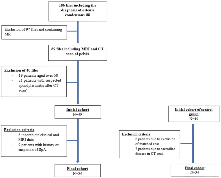
Methods: A total of 34 patients with OCI, including 29 with MRI, were recruited for retrospective analysis. The SIJ MRIs were retrospectively analysed by two readers. A sex- and age-matched control group of patients without SIJ disorders was included. In both groups, the presence of structural bone abnormalities was assessed by CT scan analysis, and socio-demographic data were obtained by telephone questionnaire. A longitudinal analysis was conducted on patients who had undergone multiple imaging examinations.
Results: All patients were female with a mean age of 34 years. BME was observed in 66% (19/29) of OCI patients. BME in OCI was mainly located in the anterior-middle quadrant (43.48%). OCI patients had significantly more than one delivery (P = 0.0094, McNemar test), even if OCI was found in four nulliparous patients (15%). OCI patients experienced significantly more pain (P = 0.0026, McNemar test).
Conclusion: OCI is an entity found in both pregnant and non-pregnant young women. SIJ BME was found in two-thirds of OCI patients. OCI is a significant cause of BME and should be carefully considered by clinicians when dealing with a patient with low back pain in order to avoid misdiagnosing spondyloarthritis in the presence of BME of the SIJ.

 求助内容:
求助内容: 应助结果提醒方式:


