{"title":"眼科学术中光学相干断层扫描:技术与应用。","authors":"Yuankai K Tao","doi":"10.4103/tjo.TJO-D-25-00076","DOIUrl":null,"url":null,"abstract":"<p><p>Intraoperative optical coherence tomography (iOCT) offers valuable real-time, depth-resolved visualization of ocular anatomy and during ophthalmic surgical maneuvers, which can be used to augment clinical decision-making, help verify surgical endpoints, enhance surgical precision, and facilitate the development of novel surgical techniques. Early iOCT demonstrations used perioperative devices, such as handheld and intraocular probes, which required pauses in surgery and disrupted clinical workflow. The advent of microscope-integrated systems addressed these limitations, allowing for iOCT imaging concurrent with surgical microscopy. iOCT image visualization has similarly progressed from external monitors, which require surgeons to divert their gaze, to heads-up displays integrated into microscope oculars, enabling direct overlays and improved ergonomics. Most recent advances have included increasing imaging speed to enable four-dimensional visualization of surgical dynamics and integration of automated surgical instrument tracking technologies. Clinical translation of iOCT has demonstrated utility across a range of procedures, including glaucoma surgery, corneal transplants, cataract extraction, vitrectomy, membrane peel, retinal detachment and macular hole repair, subretinal injection, and retinal prosthesis placement. As more advanced technologies are integrated into the conventional ophthalmic surgical workflow, iOCT has the potential to improve surgical performance and patient outcomes.</p>","PeriodicalId":44978,"journal":{"name":"Taiwan Journal of Ophthalmology","volume":"15 3","pages":"378-388"},"PeriodicalIF":1.2000,"publicationDate":"2025-09-05","publicationTypes":"Journal Article","fieldsOfStudy":null,"isOpenAccess":false,"openAccessPdf":"https://www.ncbi.nlm.nih.gov/pmc/articles/PMC12456914/pdf/","citationCount":"0","resultStr":"{\"title\":\"Intraoperative optical coherence tomography in ophthalmology: Technologies and applications.\",\"authors\":\"Yuankai K Tao\",\"doi\":\"10.4103/tjo.TJO-D-25-00076\",\"DOIUrl\":null,\"url\":null,\"abstract\":\"<p><p>Intraoperative optical coherence tomography (iOCT) offers valuable real-time, depth-resolved visualization of ocular anatomy and during ophthalmic surgical maneuvers, which can be used to augment clinical decision-making, help verify surgical endpoints, enhance surgical precision, and facilitate the development of novel surgical techniques. Early iOCT demonstrations used perioperative devices, such as handheld and intraocular probes, which required pauses in surgery and disrupted clinical workflow. The advent of microscope-integrated systems addressed these limitations, allowing for iOCT imaging concurrent with surgical microscopy. iOCT image visualization has similarly progressed from external monitors, which require surgeons to divert their gaze, to heads-up displays integrated into microscope oculars, enabling direct overlays and improved ergonomics. Most recent advances have included increasing imaging speed to enable four-dimensional visualization of surgical dynamics and integration of automated surgical instrument tracking technologies. Clinical translation of iOCT has demonstrated utility across a range of procedures, including glaucoma surgery, corneal transplants, cataract extraction, vitrectomy, membrane peel, retinal detachment and macular hole repair, subretinal injection, and retinal prosthesis placement. As more advanced technologies are integrated into the conventional ophthalmic surgical workflow, iOCT has the potential to improve surgical performance and patient outcomes.</p>\",\"PeriodicalId\":44978,\"journal\":{\"name\":\"Taiwan Journal of Ophthalmology\",\"volume\":\"15 3\",\"pages\":\"378-388\"},\"PeriodicalIF\":1.2000,\"publicationDate\":\"2025-09-05\",\"publicationTypes\":\"Journal Article\",\"fieldsOfStudy\":null,\"isOpenAccess\":false,\"openAccessPdf\":\"https://www.ncbi.nlm.nih.gov/pmc/articles/PMC12456914/pdf/\",\"citationCount\":\"0\",\"resultStr\":null,\"platform\":\"Semanticscholar\",\"paperid\":null,\"PeriodicalName\":\"Taiwan Journal of Ophthalmology\",\"FirstCategoryId\":\"1085\",\"ListUrlMain\":\"https://doi.org/10.4103/tjo.TJO-D-25-00076\",\"RegionNum\":0,\"RegionCategory\":null,\"ArticlePicture\":[],\"TitleCN\":null,\"AbstractTextCN\":null,\"PMCID\":null,\"EPubDate\":\"2025/7/1 0:00:00\",\"PubModel\":\"eCollection\",\"JCR\":\"Q4\",\"JCRName\":\"OPHTHALMOLOGY\",\"Score\":null,\"Total\":0}","platform":"Semanticscholar","paperid":null,"PeriodicalName":"Taiwan Journal of Ophthalmology","FirstCategoryId":"1085","ListUrlMain":"https://doi.org/10.4103/tjo.TJO-D-25-00076","RegionNum":0,"RegionCategory":null,"ArticlePicture":[],"TitleCN":null,"AbstractTextCN":null,"PMCID":null,"EPubDate":"2025/7/1 0:00:00","PubModel":"eCollection","JCR":"Q4","JCRName":"OPHTHALMOLOGY","Score":null,"Total":0}
Intraoperative optical coherence tomography in ophthalmology: Technologies and applications.
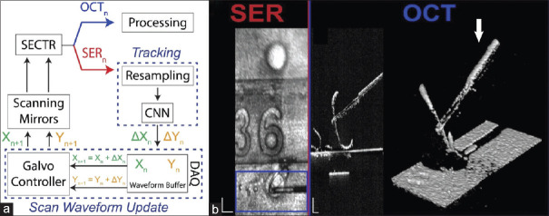
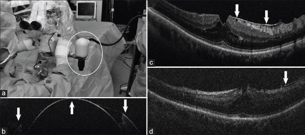
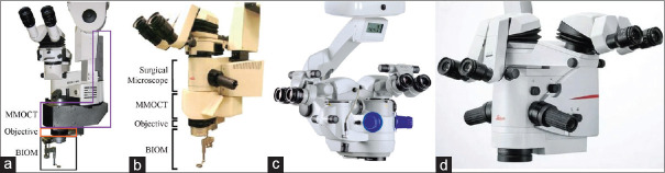
Intraoperative optical coherence tomography (iOCT) offers valuable real-time, depth-resolved visualization of ocular anatomy and during ophthalmic surgical maneuvers, which can be used to augment clinical decision-making, help verify surgical endpoints, enhance surgical precision, and facilitate the development of novel surgical techniques. Early iOCT demonstrations used perioperative devices, such as handheld and intraocular probes, which required pauses in surgery and disrupted clinical workflow. The advent of microscope-integrated systems addressed these limitations, allowing for iOCT imaging concurrent with surgical microscopy. iOCT image visualization has similarly progressed from external monitors, which require surgeons to divert their gaze, to heads-up displays integrated into microscope oculars, enabling direct overlays and improved ergonomics. Most recent advances have included increasing imaging speed to enable four-dimensional visualization of surgical dynamics and integration of automated surgical instrument tracking technologies. Clinical translation of iOCT has demonstrated utility across a range of procedures, including glaucoma surgery, corneal transplants, cataract extraction, vitrectomy, membrane peel, retinal detachment and macular hole repair, subretinal injection, and retinal prosthesis placement. As more advanced technologies are integrated into the conventional ophthalmic surgical workflow, iOCT has the potential to improve surgical performance and patient outcomes.

 求助内容:
求助内容: 应助结果提醒方式:


