Kevin Bloch-Maier, Sophie-Myriam Dridi, Arek Sulukdjian, Anne-Laure Ejeil
{"title":"小男孩颌骨黄色肉芽肿性骨髓炎1例报告。","authors":"Kevin Bloch-Maier, Sophie-Myriam Dridi, Arek Sulukdjian, Anne-Laure Ejeil","doi":"10.1186/s13023-025-04010-w","DOIUrl":null,"url":null,"abstract":"<p><p>Xanthogranulomatous inflammation is a rare chronic inflammatory condition. It has been described in the long bones, and can affect various organs, including the salivary glands, gallbladder, kidneys, and gastrointestinal tract. The diagnosis is made throught histopathology which reveales foamy macrophages alongside polymorphonuclear leukocytes, plasma cells, and polyclonal lymphocytes arranged in a mosaic-like pattern. Here, we present the case of a 16-year-old boy in good general health who was referred by his orthodontist to the oral surgery department at Bretonneau Hospital for an asymptomatic multipartite image of the left mandibular angle that was discovered incidentally on a panoramic radiograph. The clinical examination was unremarkable. Cone beam computed tomography revealed a multipartite osteolytic lesion of the left mandibular angle, but this that was not specific enough for a diagnosis. Magnetic resonance imaging ruled out a vascular malformation. A biopsy revealed xanthogranulomatous osteomyelitis. In summary, this is the second case of xanthogranulomatous osteomyelitis localised to the mandible. This highlights a crucial point : radiological images of xanthogranulomatous osteomyelitis do not allow for a diagnosis; a biopsy is essential to rule out an aggressive or a malignant tumor.</p>","PeriodicalId":19651,"journal":{"name":"Orphanet Journal of Rare Diseases","volume":"20 1","pages":"488"},"PeriodicalIF":3.5000,"publicationDate":"2025-09-24","publicationTypes":"Journal Article","fieldsOfStudy":null,"isOpenAccess":false,"openAccessPdf":"https://www.ncbi.nlm.nih.gov/pmc/articles/PMC12462011/pdf/","citationCount":"0","resultStr":"{\"title\":\"Xanthogranulomatous osteomyelitis of the jaw in a young boy: a case report.\",\"authors\":\"Kevin Bloch-Maier, Sophie-Myriam Dridi, Arek Sulukdjian, Anne-Laure Ejeil\",\"doi\":\"10.1186/s13023-025-04010-w\",\"DOIUrl\":null,\"url\":null,\"abstract\":\"<p><p>Xanthogranulomatous inflammation is a rare chronic inflammatory condition. It has been described in the long bones, and can affect various organs, including the salivary glands, gallbladder, kidneys, and gastrointestinal tract. The diagnosis is made throught histopathology which reveales foamy macrophages alongside polymorphonuclear leukocytes, plasma cells, and polyclonal lymphocytes arranged in a mosaic-like pattern. Here, we present the case of a 16-year-old boy in good general health who was referred by his orthodontist to the oral surgery department at Bretonneau Hospital for an asymptomatic multipartite image of the left mandibular angle that was discovered incidentally on a panoramic radiograph. The clinical examination was unremarkable. Cone beam computed tomography revealed a multipartite osteolytic lesion of the left mandibular angle, but this that was not specific enough for a diagnosis. Magnetic resonance imaging ruled out a vascular malformation. A biopsy revealed xanthogranulomatous osteomyelitis. In summary, this is the second case of xanthogranulomatous osteomyelitis localised to the mandible. This highlights a crucial point : radiological images of xanthogranulomatous osteomyelitis do not allow for a diagnosis; a biopsy is essential to rule out an aggressive or a malignant tumor.</p>\",\"PeriodicalId\":19651,\"journal\":{\"name\":\"Orphanet Journal of Rare Diseases\",\"volume\":\"20 1\",\"pages\":\"488\"},\"PeriodicalIF\":3.5000,\"publicationDate\":\"2025-09-24\",\"publicationTypes\":\"Journal Article\",\"fieldsOfStudy\":null,\"isOpenAccess\":false,\"openAccessPdf\":\"https://www.ncbi.nlm.nih.gov/pmc/articles/PMC12462011/pdf/\",\"citationCount\":\"0\",\"resultStr\":null,\"platform\":\"Semanticscholar\",\"paperid\":null,\"PeriodicalName\":\"Orphanet Journal of Rare Diseases\",\"FirstCategoryId\":\"3\",\"ListUrlMain\":\"https://doi.org/10.1186/s13023-025-04010-w\",\"RegionNum\":2,\"RegionCategory\":\"医学\",\"ArticlePicture\":[],\"TitleCN\":null,\"AbstractTextCN\":null,\"PMCID\":null,\"EPubDate\":\"\",\"PubModel\":\"\",\"JCR\":\"Q2\",\"JCRName\":\"GENETICS & HEREDITY\",\"Score\":null,\"Total\":0}","platform":"Semanticscholar","paperid":null,"PeriodicalName":"Orphanet Journal of Rare Diseases","FirstCategoryId":"3","ListUrlMain":"https://doi.org/10.1186/s13023-025-04010-w","RegionNum":2,"RegionCategory":"医学","ArticlePicture":[],"TitleCN":null,"AbstractTextCN":null,"PMCID":null,"EPubDate":"","PubModel":"","JCR":"Q2","JCRName":"GENETICS & HEREDITY","Score":null,"Total":0}
Xanthogranulomatous osteomyelitis of the jaw in a young boy: a case report.
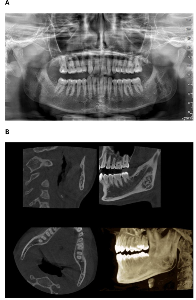
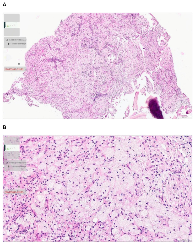
Xanthogranulomatous inflammation is a rare chronic inflammatory condition. It has been described in the long bones, and can affect various organs, including the salivary glands, gallbladder, kidneys, and gastrointestinal tract. The diagnosis is made throught histopathology which reveales foamy macrophages alongside polymorphonuclear leukocytes, plasma cells, and polyclonal lymphocytes arranged in a mosaic-like pattern. Here, we present the case of a 16-year-old boy in good general health who was referred by his orthodontist to the oral surgery department at Bretonneau Hospital for an asymptomatic multipartite image of the left mandibular angle that was discovered incidentally on a panoramic radiograph. The clinical examination was unremarkable. Cone beam computed tomography revealed a multipartite osteolytic lesion of the left mandibular angle, but this that was not specific enough for a diagnosis. Magnetic resonance imaging ruled out a vascular malformation. A biopsy revealed xanthogranulomatous osteomyelitis. In summary, this is the second case of xanthogranulomatous osteomyelitis localised to the mandible. This highlights a crucial point : radiological images of xanthogranulomatous osteomyelitis do not allow for a diagnosis; a biopsy is essential to rule out an aggressive or a malignant tumor.
期刊介绍:
Orphanet Journal of Rare Diseases is an open access, peer-reviewed journal that encompasses all aspects of rare diseases and orphan drugs. The journal publishes high-quality reviews on specific rare diseases. In addition, the journal may consider articles on clinical trial outcome reports, either positive or negative, and articles on public health issues in the field of rare diseases and orphan drugs. The journal does not accept case reports.


 求助内容:
求助内容: 应助结果提醒方式:


