Li Sun, Yaomin Wang, Kefan Zhang, Xinlong Chen, Hui Shi
{"title":"贫白细胞与富白细胞富血小板血浆治疗兔膝骨性关节炎的疗效比较及自噬机制的研究。","authors":"Li Sun, Yaomin Wang, Kefan Zhang, Xinlong Chen, Hui Shi","doi":"10.1186/s12891-025-08906-3","DOIUrl":null,"url":null,"abstract":"<p><p>Osteoarthritis of the knee (KOA) is a degenerative disease characterized by the deterioration, destruction, and proliferation of articular cartilage and synovium. Autophagy, a crucial intracellular homeostatic mechanism, significantly contributes to the pathogenesis of OA. Platelet-rich plasma (PRP), derived from autologous blood, contains a high concentration of platelets that secrete various growth factors, promoting the regeneration and repair of joint structures. In this experiment, we compared leukocyte-rich platelet-rich plasma (LR-PRP) with leukocyte-poor platelet-rich plasma (LP-PRP) in a rabbit osteoarthritis model induced by papain to investigate the therapeutic efficacy and mechanism of action of platelet-rich plasma on osteoarthritis. Additionally, we analyzed whether different leukocyte concentrations in platelet-rich plasma would affect the treatment outcomes. Papain was injected into the knee joints of 32 rabbits to induce osteoarthritis, and the animals were cultured until bone maturity. LP-PRP and LR-PRP were prepared using different centrifugation methods and injected into the knee joint cavity. Eight weeks after the injection, various analyses were performed on the rabbit knee joint synovium and cartilage. These included HE staining, immunohistochemistry for PI3K, AKT, mTOR, LC3-II, MMP-13, toluidine blue staining, and ELISA for IL-1β and TNF-α in joint fluid. The synovial membrane was scored using the Krenn score, and cartilage was evaluated with the Mankin pathology score. Quantitative analysis of immunohistochemistry was conducted using ImageJ image processing software. The rabbit osteoarthritis model was successfully established by injecting papain into the knee joint cavity. All indices showed significant differences between the model group and the control group (P < 0.05). Both LP-PRP and LR-PRP exhibited therapeutic effects compared to the model group (P < 0.05). The concentrations of IL-1β and TNF-α in the LP-PRP group were significantly lower than those in the LR-PRP group (P < 0.05). However, no significant differences were observed in the morphology of the synovial membrane, cartilage, and other indices between the LR-PRP and LP-PRP groups (P > 0.05).</p>","PeriodicalId":9189,"journal":{"name":"BMC Musculoskeletal Disorders","volume":"26 1","pages":"857"},"PeriodicalIF":2.4000,"publicationDate":"2025-09-24","publicationTypes":"Journal Article","fieldsOfStudy":null,"isOpenAccess":false,"openAccessPdf":"https://www.ncbi.nlm.nih.gov/pmc/articles/PMC12461960/pdf/","citationCount":"0","resultStr":"{\"title\":\"Comparison of the therapeutic efficacy of leukocyte-poor and leukocyte-rich platelet-rich plasma in rabbit knee osteoarthritis and the study of autophagy mechanism.\",\"authors\":\"Li Sun, Yaomin Wang, Kefan Zhang, Xinlong Chen, Hui Shi\",\"doi\":\"10.1186/s12891-025-08906-3\",\"DOIUrl\":null,\"url\":null,\"abstract\":\"<p><p>Osteoarthritis of the knee (KOA) is a degenerative disease characterized by the deterioration, destruction, and proliferation of articular cartilage and synovium. Autophagy, a crucial intracellular homeostatic mechanism, significantly contributes to the pathogenesis of OA. Platelet-rich plasma (PRP), derived from autologous blood, contains a high concentration of platelets that secrete various growth factors, promoting the regeneration and repair of joint structures. In this experiment, we compared leukocyte-rich platelet-rich plasma (LR-PRP) with leukocyte-poor platelet-rich plasma (LP-PRP) in a rabbit osteoarthritis model induced by papain to investigate the therapeutic efficacy and mechanism of action of platelet-rich plasma on osteoarthritis. Additionally, we analyzed whether different leukocyte concentrations in platelet-rich plasma would affect the treatment outcomes. Papain was injected into the knee joints of 32 rabbits to induce osteoarthritis, and the animals were cultured until bone maturity. LP-PRP and LR-PRP were prepared using different centrifugation methods and injected into the knee joint cavity. Eight weeks after the injection, various analyses were performed on the rabbit knee joint synovium and cartilage. These included HE staining, immunohistochemistry for PI3K, AKT, mTOR, LC3-II, MMP-13, toluidine blue staining, and ELISA for IL-1β and TNF-α in joint fluid. The synovial membrane was scored using the Krenn score, and cartilage was evaluated with the Mankin pathology score. Quantitative analysis of immunohistochemistry was conducted using ImageJ image processing software. The rabbit osteoarthritis model was successfully established by injecting papain into the knee joint cavity. All indices showed significant differences between the model group and the control group (P < 0.05). Both LP-PRP and LR-PRP exhibited therapeutic effects compared to the model group (P < 0.05). The concentrations of IL-1β and TNF-α in the LP-PRP group were significantly lower than those in the LR-PRP group (P < 0.05). However, no significant differences were observed in the morphology of the synovial membrane, cartilage, and other indices between the LR-PRP and LP-PRP groups (P > 0.05).</p>\",\"PeriodicalId\":9189,\"journal\":{\"name\":\"BMC Musculoskeletal Disorders\",\"volume\":\"26 1\",\"pages\":\"857\"},\"PeriodicalIF\":2.4000,\"publicationDate\":\"2025-09-24\",\"publicationTypes\":\"Journal Article\",\"fieldsOfStudy\":null,\"isOpenAccess\":false,\"openAccessPdf\":\"https://www.ncbi.nlm.nih.gov/pmc/articles/PMC12461960/pdf/\",\"citationCount\":\"0\",\"resultStr\":null,\"platform\":\"Semanticscholar\",\"paperid\":null,\"PeriodicalName\":\"BMC Musculoskeletal Disorders\",\"FirstCategoryId\":\"3\",\"ListUrlMain\":\"https://doi.org/10.1186/s12891-025-08906-3\",\"RegionNum\":3,\"RegionCategory\":\"医学\",\"ArticlePicture\":[],\"TitleCN\":null,\"AbstractTextCN\":null,\"PMCID\":null,\"EPubDate\":\"\",\"PubModel\":\"\",\"JCR\":\"Q2\",\"JCRName\":\"ORTHOPEDICS\",\"Score\":null,\"Total\":0}","platform":"Semanticscholar","paperid":null,"PeriodicalName":"BMC Musculoskeletal Disorders","FirstCategoryId":"3","ListUrlMain":"https://doi.org/10.1186/s12891-025-08906-3","RegionNum":3,"RegionCategory":"医学","ArticlePicture":[],"TitleCN":null,"AbstractTextCN":null,"PMCID":null,"EPubDate":"","PubModel":"","JCR":"Q2","JCRName":"ORTHOPEDICS","Score":null,"Total":0}
Comparison of the therapeutic efficacy of leukocyte-poor and leukocyte-rich platelet-rich plasma in rabbit knee osteoarthritis and the study of autophagy mechanism.
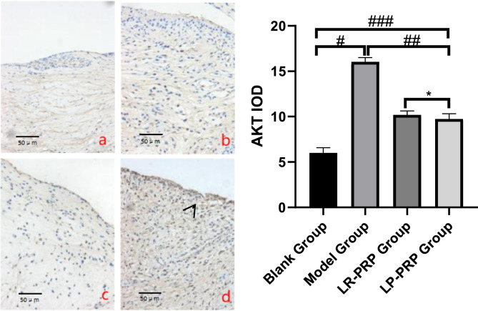
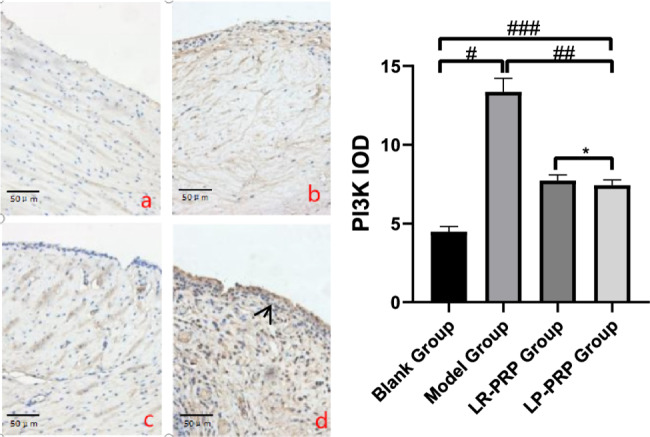
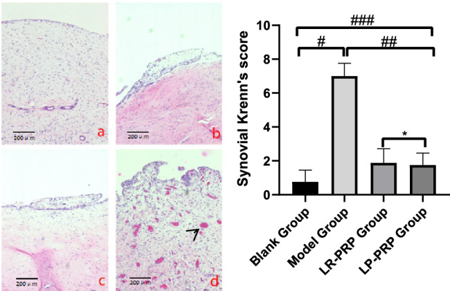
Osteoarthritis of the knee (KOA) is a degenerative disease characterized by the deterioration, destruction, and proliferation of articular cartilage and synovium. Autophagy, a crucial intracellular homeostatic mechanism, significantly contributes to the pathogenesis of OA. Platelet-rich plasma (PRP), derived from autologous blood, contains a high concentration of platelets that secrete various growth factors, promoting the regeneration and repair of joint structures. In this experiment, we compared leukocyte-rich platelet-rich plasma (LR-PRP) with leukocyte-poor platelet-rich plasma (LP-PRP) in a rabbit osteoarthritis model induced by papain to investigate the therapeutic efficacy and mechanism of action of platelet-rich plasma on osteoarthritis. Additionally, we analyzed whether different leukocyte concentrations in platelet-rich plasma would affect the treatment outcomes. Papain was injected into the knee joints of 32 rabbits to induce osteoarthritis, and the animals were cultured until bone maturity. LP-PRP and LR-PRP were prepared using different centrifugation methods and injected into the knee joint cavity. Eight weeks after the injection, various analyses were performed on the rabbit knee joint synovium and cartilage. These included HE staining, immunohistochemistry for PI3K, AKT, mTOR, LC3-II, MMP-13, toluidine blue staining, and ELISA for IL-1β and TNF-α in joint fluid. The synovial membrane was scored using the Krenn score, and cartilage was evaluated with the Mankin pathology score. Quantitative analysis of immunohistochemistry was conducted using ImageJ image processing software. The rabbit osteoarthritis model was successfully established by injecting papain into the knee joint cavity. All indices showed significant differences between the model group and the control group (P < 0.05). Both LP-PRP and LR-PRP exhibited therapeutic effects compared to the model group (P < 0.05). The concentrations of IL-1β and TNF-α in the LP-PRP group were significantly lower than those in the LR-PRP group (P < 0.05). However, no significant differences were observed in the morphology of the synovial membrane, cartilage, and other indices between the LR-PRP and LP-PRP groups (P > 0.05).
期刊介绍:
BMC Musculoskeletal Disorders is an open access, peer-reviewed journal that considers articles on all aspects of the prevention, diagnosis and management of musculoskeletal disorders, as well as related molecular genetics, pathophysiology, and epidemiology.
The scope of the Journal covers research into rheumatic diseases where the primary focus relates specifically to a component(s) of the musculoskeletal system.
 求助内容:
求助内容: 应助结果提醒方式:


