Iván Andrés Pineda-Betancurt, Luis Ernesto Ballesteros-Acuña, Fabian Alejandro Gömez-Torres
{"title":"犬冠状窦的直接解剖特征(犬狼疮)。","authors":"Iván Andrés Pineda-Betancurt, Luis Ernesto Ballesteros-Acuña, Fabian Alejandro Gömez-Torres","doi":"10.5455/OVJ.2025.v15.i6.53","DOIUrl":null,"url":null,"abstract":"<p><strong>Background: </strong>The inquiry on the venous drainage of the heart of dogs is still under development, despite its significant importance in the framework of comparative anatomy and its implications for veterinary medicine.</p><p><strong>Aim: </strong>The objective of the study was to perform a qualitative and biometric characterization of the cardiac veins of dogs.</p><p><strong>Methods: </strong>Using the venous bed perfusion technique, semi-synthetic resin (Palatal GP40L 80% and styrene 20% with blue dye) was injected into the venous system of 32 hearts of dogs that died in veterinary clinics, categorized by weight as follows: <10 kg; 10-19 kg; 20-29 kg; ≥30 Kg.</p><p><strong>Results: </strong>The length and diameter of the coronary sinus for the <10 kg group were 18.49 ± 3.31 and 4.40 ± 0.85 mm. Its morphology was cylindrical in 50% of cases and funnel-shaped in 34.4% of them. Arteriovenous trigone was observed in 93.8% of hearts, with a prevalence of closed configuration in 60% of cases, in both the superior and inferior segments. The diameter of the great cardiac vein at the level of the paraconal interventricular sulcus for the 20-29 kg group was 2.13 ± 0.49 mm, while its distal diameter was 4.03 ± 0.32 mm. The left marginal vein originated mainly in the lower third of the left margin (46.9%), and its caliber at the drainage (2.84 mm) was larger in cases at the level of the coronary sinus than in the great cardiac vein (<i>p</i> = 0.012). Venous anastomoses between the middle and great cardiac veins were observed in 50% of the hearts.</p><p><strong>Conclusion: </strong>The qualitative and biometric characteristics of the venous structures of the heart of dogs were described in detail and categorized by weight groups of the animals. These results are highly useful for the implementation of endovascular devices and the treatment of cardiac diseases.</p>","PeriodicalId":19531,"journal":{"name":"Open Veterinary Journal","volume":"15 6","pages":"2840-2848"},"PeriodicalIF":1.0000,"publicationDate":"2025-06-01","publicationTypes":"Journal Article","fieldsOfStudy":null,"isOpenAccess":false,"openAccessPdf":"https://www.ncbi.nlm.nih.gov/pmc/articles/PMC12451137/pdf/","citationCount":"0","resultStr":"{\"title\":\"A direct anatomical characterization of the coronary sinus in dogs (<i>Canis lupus</i> familiaris).\",\"authors\":\"Iván Andrés Pineda-Betancurt, Luis Ernesto Ballesteros-Acuña, Fabian Alejandro Gömez-Torres\",\"doi\":\"10.5455/OVJ.2025.v15.i6.53\",\"DOIUrl\":null,\"url\":null,\"abstract\":\"<p><strong>Background: </strong>The inquiry on the venous drainage of the heart of dogs is still under development, despite its significant importance in the framework of comparative anatomy and its implications for veterinary medicine.</p><p><strong>Aim: </strong>The objective of the study was to perform a qualitative and biometric characterization of the cardiac veins of dogs.</p><p><strong>Methods: </strong>Using the venous bed perfusion technique, semi-synthetic resin (Palatal GP40L 80% and styrene 20% with blue dye) was injected into the venous system of 32 hearts of dogs that died in veterinary clinics, categorized by weight as follows: <10 kg; 10-19 kg; 20-29 kg; ≥30 Kg.</p><p><strong>Results: </strong>The length and diameter of the coronary sinus for the <10 kg group were 18.49 ± 3.31 and 4.40 ± 0.85 mm. Its morphology was cylindrical in 50% of cases and funnel-shaped in 34.4% of them. Arteriovenous trigone was observed in 93.8% of hearts, with a prevalence of closed configuration in 60% of cases, in both the superior and inferior segments. The diameter of the great cardiac vein at the level of the paraconal interventricular sulcus for the 20-29 kg group was 2.13 ± 0.49 mm, while its distal diameter was 4.03 ± 0.32 mm. The left marginal vein originated mainly in the lower third of the left margin (46.9%), and its caliber at the drainage (2.84 mm) was larger in cases at the level of the coronary sinus than in the great cardiac vein (<i>p</i> = 0.012). Venous anastomoses between the middle and great cardiac veins were observed in 50% of the hearts.</p><p><strong>Conclusion: </strong>The qualitative and biometric characteristics of the venous structures of the heart of dogs were described in detail and categorized by weight groups of the animals. These results are highly useful for the implementation of endovascular devices and the treatment of cardiac diseases.</p>\",\"PeriodicalId\":19531,\"journal\":{\"name\":\"Open Veterinary Journal\",\"volume\":\"15 6\",\"pages\":\"2840-2848\"},\"PeriodicalIF\":1.0000,\"publicationDate\":\"2025-06-01\",\"publicationTypes\":\"Journal Article\",\"fieldsOfStudy\":null,\"isOpenAccess\":false,\"openAccessPdf\":\"https://www.ncbi.nlm.nih.gov/pmc/articles/PMC12451137/pdf/\",\"citationCount\":\"0\",\"resultStr\":null,\"platform\":\"Semanticscholar\",\"paperid\":null,\"PeriodicalName\":\"Open Veterinary Journal\",\"FirstCategoryId\":\"1085\",\"ListUrlMain\":\"https://doi.org/10.5455/OVJ.2025.v15.i6.53\",\"RegionNum\":0,\"RegionCategory\":null,\"ArticlePicture\":[],\"TitleCN\":null,\"AbstractTextCN\":null,\"PMCID\":null,\"EPubDate\":\"2025/6/30 0:00:00\",\"PubModel\":\"Epub\",\"JCR\":\"Q3\",\"JCRName\":\"VETERINARY SCIENCES\",\"Score\":null,\"Total\":0}","platform":"Semanticscholar","paperid":null,"PeriodicalName":"Open Veterinary Journal","FirstCategoryId":"1085","ListUrlMain":"https://doi.org/10.5455/OVJ.2025.v15.i6.53","RegionNum":0,"RegionCategory":null,"ArticlePicture":[],"TitleCN":null,"AbstractTextCN":null,"PMCID":null,"EPubDate":"2025/6/30 0:00:00","PubModel":"Epub","JCR":"Q3","JCRName":"VETERINARY SCIENCES","Score":null,"Total":0}
A direct anatomical characterization of the coronary sinus in dogs (Canis lupus familiaris).
Background: The inquiry on the venous drainage of the heart of dogs is still under development, despite its significant importance in the framework of comparative anatomy and its implications for veterinary medicine.
Aim: The objective of the study was to perform a qualitative and biometric characterization of the cardiac veins of dogs.
Methods: Using the venous bed perfusion technique, semi-synthetic resin (Palatal GP40L 80% and styrene 20% with blue dye) was injected into the venous system of 32 hearts of dogs that died in veterinary clinics, categorized by weight as follows: <10 kg; 10-19 kg; 20-29 kg; ≥30 Kg.
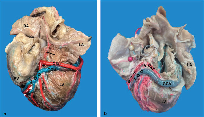
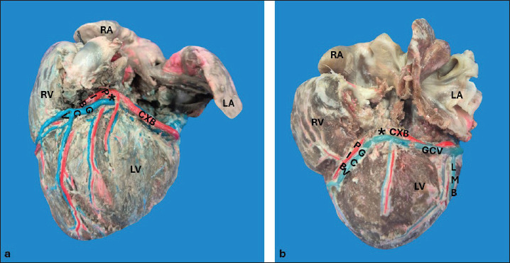
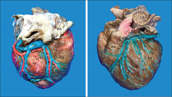
Results: The length and diameter of the coronary sinus for the <10 kg group were 18.49 ± 3.31 and 4.40 ± 0.85 mm. Its morphology was cylindrical in 50% of cases and funnel-shaped in 34.4% of them. Arteriovenous trigone was observed in 93.8% of hearts, with a prevalence of closed configuration in 60% of cases, in both the superior and inferior segments. The diameter of the great cardiac vein at the level of the paraconal interventricular sulcus for the 20-29 kg group was 2.13 ± 0.49 mm, while its distal diameter was 4.03 ± 0.32 mm. The left marginal vein originated mainly in the lower third of the left margin (46.9%), and its caliber at the drainage (2.84 mm) was larger in cases at the level of the coronary sinus than in the great cardiac vein (p = 0.012). Venous anastomoses between the middle and great cardiac veins were observed in 50% of the hearts.
Conclusion: The qualitative and biometric characteristics of the venous structures of the heart of dogs were described in detail and categorized by weight groups of the animals. These results are highly useful for the implementation of endovascular devices and the treatment of cardiac diseases.
期刊介绍:
Open Veterinary Journal is a peer-reviewed international open access online and printed journal that publishes high-quality original research articles. reviews, short communications and case reports dedicated to all aspects of veterinary sciences and its related subjects. Research areas include the following: Infectious diseases of zoonotic/food-borne importance, applied biochemistry, parasitology, endocrinology, microbiology, immunology, pathology, pharmacology, physiology, epidemiology, molecular biology, immunogenetics, surgery, ophthalmology, dermatology, oncology and animal reproduction. All papers are peer-reviewed. Moreover, with the presence of well-qualified group of international referees, the process of publication will be done meticulously and to the highest standards.

 求助内容:
求助内容: 应助结果提醒方式:


