使用倾斜校正扫描透射电子显微镜对厚样品进行剂量有效的冷冻电子显微镜。
IF 32.1
1区 生物学
Q1 BIOCHEMICAL RESEARCH METHODS
引用次数: 0
摘要
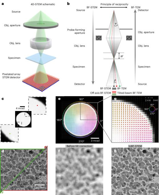
低温电子显微镜是结构生物学的有力工具。在厚的样品中,挑战出现了,因为透射电子的一个指数大的分数失去能量,从非弹性散射和不能再正确聚焦的结果,色差在样品后光学。而不是以降低电位信号为代价过滤掉非弹性散射,正如在能量过滤透射电子显微镜中所做的那样,我们展示了如何使用倾斜校正的亮场扫描透射电子显微镜数据在像素化检测器上收集快速获得剂量有效和未过滤的图像。与能量过滤透射电镜相比,使用倾斜校正的亮场扫描透射电镜对厚度超过500 nm的完整细菌细胞和大型细胞器的二维图像进行对比度增强,剂量效率提高3-5倍。为了证明该技术在结构确定方面的性能,我们提出了一个亚纳米分辨率的单颗粒分析图,用于从789个颗粒中确定高度对称的病毒样颗粒。本文章由计算机程序翻译,如有差异,请以英文原文为准。
Dose-efficient cryo-electron microscopy for thick samples using tilt- corrected scanning transmission electron microscopy
Cryogenic electron microscopy is a powerful tool in structural biology. In thick specimens, challenges arise as an exponentially larger fraction of the transmitted electrons lose energy from inelastic scattering and can no longer be properly focused as a result of chromatic aberrations in the post-specimen optics. Rather than filtering out the inelastic scattering at the price of reducing potential signal, as is done in energy-filtered transmission electron microscopy, we show how a dose-efficient and unfiltered image can be rapidly obtained using tilt-corrected bright-field scanning transmission electron microscopy data collected on a pixelated detector. Enhanced contrast and a 3–5× improvement in dose efficiency are observed for two-dimensional images of intact bacterial cells and large organelles using tilt-corrected bright-field scanning transmission electron microscopy compared to energy-filtered transmission electron microscopy for thicknesses beyond 500 nm. As a proof of concept for the technique’s performance in structural determination, we present a single-particle analysis map at sub-nanometer resolution for a highly symmetric virus-like particle determined from 789 particles. Tilt-corrected bright-field scanning transmission electron microscopy offers enhanced cryogenic electron microscopy contrast and substantial improvement in dose efficiency for thick samples such as bacterial cells and large organelles, while still being able to perform single-particle analysis.
求助全文
通过发布文献求助,成功后即可免费获取论文全文。
去求助
来源期刊
Nature Methods
生物-生化研究方法
CiteScore
58.70
自引率
1.70%
发文量
326
审稿时长
1 months
期刊介绍:
Nature Methods is a monthly journal that focuses on publishing innovative methods and substantial enhancements to fundamental life sciences research techniques. Geared towards a diverse, interdisciplinary readership of researchers in academia and industry engaged in laboratory work, the journal offers new tools for research and emphasizes the immediate practical significance of the featured work. It publishes primary research papers and reviews recent technical and methodological advancements, with a particular interest in primary methods papers relevant to the biological and biomedical sciences. This includes methods rooted in chemistry with practical applications for studying biological problems.
 求助内容:
求助内容: 应助结果提醒方式:


