{"title":"视频耳镜辅助钬:钇铝石榴石激光切除4只犬的耳部肿物。","authors":"Adheip Prabakaran, Julia P Sumner, Sophie A Tyler","doi":"10.5455/OVJ.2025.v15.i6.61","DOIUrl":null,"url":null,"abstract":"<p><strong>Background: </strong>This case series reports the outcomes of four dogs that underwent video otoscopy-assisted laser excision (VALE) of aural masses using a Holmium: yttrium-aluminum-garnet (Ho:YAG) laser.</p><p><strong>Case description: </strong>All cases underwent excision of aural masses by snare polypectomy and laser ablation of the stalk or direct excision of the mass with a Ho:YAG laser. The first three cases underwent VALE and were free of clinical signs at follow-up times ranging from 197 to 403 days post procedure. In one case, the direct application of the laser on the mass resulted in thermal necrosis of the submitted tissue and an open histopathological diagnosis. In the fourth case, VALE was unsuccessful due to an intimate adhesion of the mass with the tympanic bulla. This patient underwent total ear canal ablation and lateral bulla osteotomy (TECALBO), which subsequently developed a deep surgical site infection and was revised. The patient experienced complete recovery at 232 days post TECALBO.</p><p><strong>Conclusion: </strong>VALE of aural masses using a Ho:YAG laser is a minimally invasive and effective treatment for aural masses in dogs in the medium term, providing an alternative to TECALBO in three out of four of our cases. Direct excision of the mass using a laser can cause thermal necrosis of the tissue, making histopathologic diagnosis more challenging. More studies are required to determine the contraindications of this technique and to better quantify the outcomes and complications.</p>","PeriodicalId":19531,"journal":{"name":"Open Veterinary Journal","volume":"15 6","pages":"2909-2914"},"PeriodicalIF":1.0000,"publicationDate":"2025-06-01","publicationTypes":"Journal Article","fieldsOfStudy":null,"isOpenAccess":false,"openAccessPdf":"https://www.ncbi.nlm.nih.gov/pmc/articles/PMC12451145/pdf/","citationCount":"0","resultStr":"{\"title\":\"Video otoscopy-assisted Holmium: yttrium-aluminum- garnet laser excision of aural masses in four dogs.\",\"authors\":\"Adheip Prabakaran, Julia P Sumner, Sophie A Tyler\",\"doi\":\"10.5455/OVJ.2025.v15.i6.61\",\"DOIUrl\":null,\"url\":null,\"abstract\":\"<p><strong>Background: </strong>This case series reports the outcomes of four dogs that underwent video otoscopy-assisted laser excision (VALE) of aural masses using a Holmium: yttrium-aluminum-garnet (Ho:YAG) laser.</p><p><strong>Case description: </strong>All cases underwent excision of aural masses by snare polypectomy and laser ablation of the stalk or direct excision of the mass with a Ho:YAG laser. The first three cases underwent VALE and were free of clinical signs at follow-up times ranging from 197 to 403 days post procedure. In one case, the direct application of the laser on the mass resulted in thermal necrosis of the submitted tissue and an open histopathological diagnosis. In the fourth case, VALE was unsuccessful due to an intimate adhesion of the mass with the tympanic bulla. This patient underwent total ear canal ablation and lateral bulla osteotomy (TECALBO), which subsequently developed a deep surgical site infection and was revised. The patient experienced complete recovery at 232 days post TECALBO.</p><p><strong>Conclusion: </strong>VALE of aural masses using a Ho:YAG laser is a minimally invasive and effective treatment for aural masses in dogs in the medium term, providing an alternative to TECALBO in three out of four of our cases. Direct excision of the mass using a laser can cause thermal necrosis of the tissue, making histopathologic diagnosis more challenging. More studies are required to determine the contraindications of this technique and to better quantify the outcomes and complications.</p>\",\"PeriodicalId\":19531,\"journal\":{\"name\":\"Open Veterinary Journal\",\"volume\":\"15 6\",\"pages\":\"2909-2914\"},\"PeriodicalIF\":1.0000,\"publicationDate\":\"2025-06-01\",\"publicationTypes\":\"Journal Article\",\"fieldsOfStudy\":null,\"isOpenAccess\":false,\"openAccessPdf\":\"https://www.ncbi.nlm.nih.gov/pmc/articles/PMC12451145/pdf/\",\"citationCount\":\"0\",\"resultStr\":null,\"platform\":\"Semanticscholar\",\"paperid\":null,\"PeriodicalName\":\"Open Veterinary Journal\",\"FirstCategoryId\":\"1085\",\"ListUrlMain\":\"https://doi.org/10.5455/OVJ.2025.v15.i6.61\",\"RegionNum\":0,\"RegionCategory\":null,\"ArticlePicture\":[],\"TitleCN\":null,\"AbstractTextCN\":null,\"PMCID\":null,\"EPubDate\":\"2025/6/30 0:00:00\",\"PubModel\":\"Epub\",\"JCR\":\"Q3\",\"JCRName\":\"VETERINARY SCIENCES\",\"Score\":null,\"Total\":0}","platform":"Semanticscholar","paperid":null,"PeriodicalName":"Open Veterinary Journal","FirstCategoryId":"1085","ListUrlMain":"https://doi.org/10.5455/OVJ.2025.v15.i6.61","RegionNum":0,"RegionCategory":null,"ArticlePicture":[],"TitleCN":null,"AbstractTextCN":null,"PMCID":null,"EPubDate":"2025/6/30 0:00:00","PubModel":"Epub","JCR":"Q3","JCRName":"VETERINARY SCIENCES","Score":null,"Total":0}
Video otoscopy-assisted Holmium: yttrium-aluminum- garnet laser excision of aural masses in four dogs.
Background: This case series reports the outcomes of four dogs that underwent video otoscopy-assisted laser excision (VALE) of aural masses using a Holmium: yttrium-aluminum-garnet (Ho:YAG) laser.
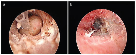
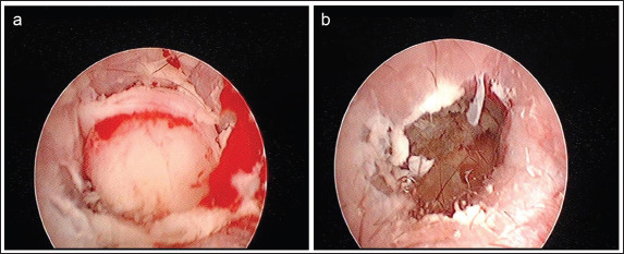
Case description: All cases underwent excision of aural masses by snare polypectomy and laser ablation of the stalk or direct excision of the mass with a Ho:YAG laser. The first three cases underwent VALE and were free of clinical signs at follow-up times ranging from 197 to 403 days post procedure. In one case, the direct application of the laser on the mass resulted in thermal necrosis of the submitted tissue and an open histopathological diagnosis. In the fourth case, VALE was unsuccessful due to an intimate adhesion of the mass with the tympanic bulla. This patient underwent total ear canal ablation and lateral bulla osteotomy (TECALBO), which subsequently developed a deep surgical site infection and was revised. The patient experienced complete recovery at 232 days post TECALBO.
Conclusion: VALE of aural masses using a Ho:YAG laser is a minimally invasive and effective treatment for aural masses in dogs in the medium term, providing an alternative to TECALBO in three out of four of our cases. Direct excision of the mass using a laser can cause thermal necrosis of the tissue, making histopathologic diagnosis more challenging. More studies are required to determine the contraindications of this technique and to better quantify the outcomes and complications.
期刊介绍:
Open Veterinary Journal is a peer-reviewed international open access online and printed journal that publishes high-quality original research articles. reviews, short communications and case reports dedicated to all aspects of veterinary sciences and its related subjects. Research areas include the following: Infectious diseases of zoonotic/food-borne importance, applied biochemistry, parasitology, endocrinology, microbiology, immunology, pathology, pharmacology, physiology, epidemiology, molecular biology, immunogenetics, surgery, ophthalmology, dermatology, oncology and animal reproduction. All papers are peer-reviewed. Moreover, with the presence of well-qualified group of international referees, the process of publication will be done meticulously and to the highest standards.

 求助内容:
求助内容: 应助结果提醒方式:


