{"title":"微针辅助局部外泌体治疗表皮和DEJ重塑的初步组织学证据:单受试者病例报告。","authors":"Young Seob Lee","doi":"10.2147/CCID.S542022","DOIUrl":null,"url":null,"abstract":"<p><strong>Purpose: </strong>This study aimed to evaluate the histological effects of microneedling-assisted topical exosome therapy on the dermoepidermal junction (DEJ) and surrounding epidermal structures in aging skin.</p><p><strong>Patients and methods: </strong>In this single-subject case report, a healthy 63-year-old male with no known dermatologic or systemic conditions underwent a single session of 0.3 mm microneedling immediately followed by topical application of an exosome-based formulation (E-50 Skin Booster, PrimaCure Co., Ltd., Korea). A 3 mm punch biopsy was collected from the postauricular area at baseline, and a follow-up biopsy was obtained eight weeks post-treatment from an adjacent site. Hematoxylin and eosin (H&E) staining was performed, and histomorphometric parameters were analyzed using high-resolution imaging and AI-assisted quantitative tools.</p><p><strong>Results: </strong>Histological analysis revealed significant remodeling of the DEJ, with a 17.08% increase in rete ridge count (<i>p</i> = 0.02) and a 7.75% increase in ridge amplitude (<i>p</i> = 0.04). Mean epidermal thickness increased by 52.33% (<i>p</i> = 0.01), and basal keratinocyte density rose by 60.90% (<i>p</i> = 0.01), indicating enhanced regenerative activity. Collagen fiber density in the upper dermis improved by 5.77% (<i>p</i> = 0.05). No adverse effects were reported, and post-treatment tissue showed no signs of inflammation or fibrosis.</p><p><strong>Conclusion: </strong>Microneedling-assisted topical exosome therapy produced notable structural improvements at the epidermal and dermoepidermal levels in aging skin. Restoration of rete ridges, enhanced keratinocyte proliferation, and increased collagen density suggest a promising role for this combined approach in skin rejuvenation. These findings are specific to microneedling-assisted topical delivery and should not be interpreted as supporting the use of injectable exosomes, which were not evaluated in this study.</p>","PeriodicalId":10447,"journal":{"name":"Clinical, Cosmetic and Investigational Dermatology","volume":"18 ","pages":"2377-2385"},"PeriodicalIF":2.2000,"publicationDate":"2025-09-18","publicationTypes":"Journal Article","fieldsOfStudy":null,"isOpenAccess":false,"openAccessPdf":"https://www.ncbi.nlm.nih.gov/pmc/articles/PMC12452975/pdf/","citationCount":"0","resultStr":"{\"title\":\"Preliminary Histological Evidence of Epidermal and DEJ Remodeling with Microneedling-Assisted Topical Exosome Therapy: A Single-Subject Case Report.\",\"authors\":\"Young Seob Lee\",\"doi\":\"10.2147/CCID.S542022\",\"DOIUrl\":null,\"url\":null,\"abstract\":\"<p><strong>Purpose: </strong>This study aimed to evaluate the histological effects of microneedling-assisted topical exosome therapy on the dermoepidermal junction (DEJ) and surrounding epidermal structures in aging skin.</p><p><strong>Patients and methods: </strong>In this single-subject case report, a healthy 63-year-old male with no known dermatologic or systemic conditions underwent a single session of 0.3 mm microneedling immediately followed by topical application of an exosome-based formulation (E-50 Skin Booster, PrimaCure Co., Ltd., Korea). A 3 mm punch biopsy was collected from the postauricular area at baseline, and a follow-up biopsy was obtained eight weeks post-treatment from an adjacent site. Hematoxylin and eosin (H&E) staining was performed, and histomorphometric parameters were analyzed using high-resolution imaging and AI-assisted quantitative tools.</p><p><strong>Results: </strong>Histological analysis revealed significant remodeling of the DEJ, with a 17.08% increase in rete ridge count (<i>p</i> = 0.02) and a 7.75% increase in ridge amplitude (<i>p</i> = 0.04). Mean epidermal thickness increased by 52.33% (<i>p</i> = 0.01), and basal keratinocyte density rose by 60.90% (<i>p</i> = 0.01), indicating enhanced regenerative activity. Collagen fiber density in the upper dermis improved by 5.77% (<i>p</i> = 0.05). No adverse effects were reported, and post-treatment tissue showed no signs of inflammation or fibrosis.</p><p><strong>Conclusion: </strong>Microneedling-assisted topical exosome therapy produced notable structural improvements at the epidermal and dermoepidermal levels in aging skin. Restoration of rete ridges, enhanced keratinocyte proliferation, and increased collagen density suggest a promising role for this combined approach in skin rejuvenation. These findings are specific to microneedling-assisted topical delivery and should not be interpreted as supporting the use of injectable exosomes, which were not evaluated in this study.</p>\",\"PeriodicalId\":10447,\"journal\":{\"name\":\"Clinical, Cosmetic and Investigational Dermatology\",\"volume\":\"18 \",\"pages\":\"2377-2385\"},\"PeriodicalIF\":2.2000,\"publicationDate\":\"2025-09-18\",\"publicationTypes\":\"Journal Article\",\"fieldsOfStudy\":null,\"isOpenAccess\":false,\"openAccessPdf\":\"https://www.ncbi.nlm.nih.gov/pmc/articles/PMC12452975/pdf/\",\"citationCount\":\"0\",\"resultStr\":null,\"platform\":\"Semanticscholar\",\"paperid\":null,\"PeriodicalName\":\"Clinical, Cosmetic and Investigational Dermatology\",\"FirstCategoryId\":\"3\",\"ListUrlMain\":\"https://doi.org/10.2147/CCID.S542022\",\"RegionNum\":4,\"RegionCategory\":\"医学\",\"ArticlePicture\":[],\"TitleCN\":null,\"AbstractTextCN\":null,\"PMCID\":null,\"EPubDate\":\"2025/1/1 0:00:00\",\"PubModel\":\"eCollection\",\"JCR\":\"Q3\",\"JCRName\":\"DERMATOLOGY\",\"Score\":null,\"Total\":0}","platform":"Semanticscholar","paperid":null,"PeriodicalName":"Clinical, Cosmetic and Investigational Dermatology","FirstCategoryId":"3","ListUrlMain":"https://doi.org/10.2147/CCID.S542022","RegionNum":4,"RegionCategory":"医学","ArticlePicture":[],"TitleCN":null,"AbstractTextCN":null,"PMCID":null,"EPubDate":"2025/1/1 0:00:00","PubModel":"eCollection","JCR":"Q3","JCRName":"DERMATOLOGY","Score":null,"Total":0}
Preliminary Histological Evidence of Epidermal and DEJ Remodeling with Microneedling-Assisted Topical Exosome Therapy: A Single-Subject Case Report.
Purpose: This study aimed to evaluate the histological effects of microneedling-assisted topical exosome therapy on the dermoepidermal junction (DEJ) and surrounding epidermal structures in aging skin.
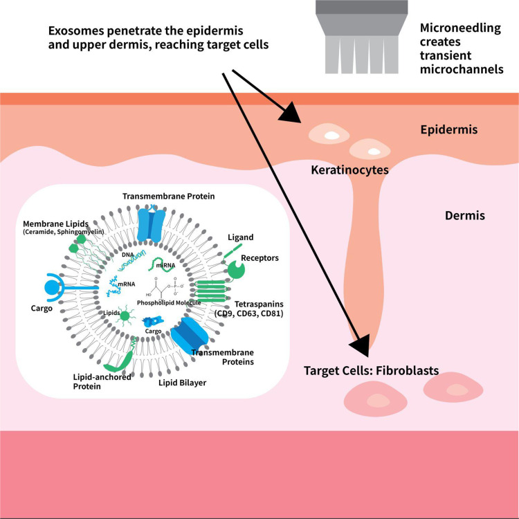
Patients and methods: In this single-subject case report, a healthy 63-year-old male with no known dermatologic or systemic conditions underwent a single session of 0.3 mm microneedling immediately followed by topical application of an exosome-based formulation (E-50 Skin Booster, PrimaCure Co., Ltd., Korea). A 3 mm punch biopsy was collected from the postauricular area at baseline, and a follow-up biopsy was obtained eight weeks post-treatment from an adjacent site. Hematoxylin and eosin (H&E) staining was performed, and histomorphometric parameters were analyzed using high-resolution imaging and AI-assisted quantitative tools.
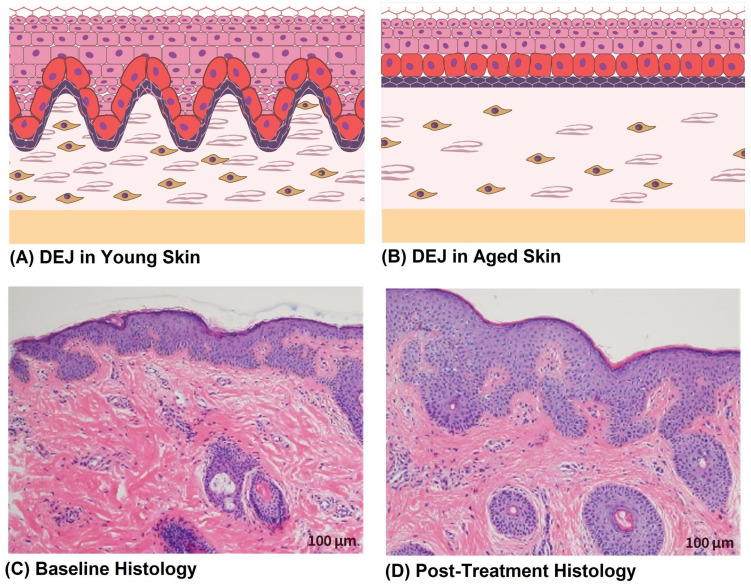
Results: Histological analysis revealed significant remodeling of the DEJ, with a 17.08% increase in rete ridge count (p = 0.02) and a 7.75% increase in ridge amplitude (p = 0.04). Mean epidermal thickness increased by 52.33% (p = 0.01), and basal keratinocyte density rose by 60.90% (p = 0.01), indicating enhanced regenerative activity. Collagen fiber density in the upper dermis improved by 5.77% (p = 0.05). No adverse effects were reported, and post-treatment tissue showed no signs of inflammation or fibrosis.
Conclusion: Microneedling-assisted topical exosome therapy produced notable structural improvements at the epidermal and dermoepidermal levels in aging skin. Restoration of rete ridges, enhanced keratinocyte proliferation, and increased collagen density suggest a promising role for this combined approach in skin rejuvenation. These findings are specific to microneedling-assisted topical delivery and should not be interpreted as supporting the use of injectable exosomes, which were not evaluated in this study.
期刊介绍:
Clinical, Cosmetic and Investigational Dermatology is an international, peer-reviewed, open access journal that focuses on the latest clinical and experimental research in all aspects of skin disease and cosmetic interventions. Normal and pathological processes in skin development and aging, their modification and treatment, as well as basic research into histology of dermal and dermal structures that provide clinical insights and potential treatment options are key topics for the journal.
Patient satisfaction, preference, quality of life, compliance, persistence and their role in developing new management options to optimize outcomes for target conditions constitute major areas of interest.
The journal is characterized by the rapid reporting of clinical studies, reviews and original research in skin research and skin care.
All areas of dermatology will be covered; contributions will be welcomed from all clinicians and basic science researchers globally.
 求助内容:
求助内容: 应助结果提醒方式:


