Néstor Porras, Lidia Sánchez-Morales, Marta Pérez-Sancho, Lucas Domínguez, Antonio Rodríguez-Bertos
{"title":"k18-hACE2小鼠与SARS-CoV-2脑感染相关的原发性小脑膜组织细胞淋巴细胞增生性疾病1例报告","authors":"Néstor Porras, Lidia Sánchez-Morales, Marta Pérez-Sancho, Lucas Domínguez, Antonio Rodríguez-Bertos","doi":"10.1186/s42826-025-00256-4","DOIUrl":null,"url":null,"abstract":"<p><strong>Background: </strong>Histiocytic proliferative disorders in the central nervous system are rare, and their potential association with viral infections remains largely unexplored. This case is relevant because it suggests a potential interaction between SARS-CoV-2 neuroinvasion and tumor development, providing insights into how viral infections might influence oncogenesis.</p><p><strong>Case presentation: </strong>A 4.5-month-old male k18-hACE-2 mouse, part of an experimental study of SARS-CoV-2, displayed a small mass in leptomeningeal area composed by neoplastic round cells. This process is associated with typical acute inflammatory and neurodegenerative lesions according to viral neuroinvasion. Histopathology revealed a well-demarcated tumor composed of lymphoblasts and intermixed with abundant histiocytic-like cells. Immunohistochemistry showed high expression of Iba-1 in histiocytes but negative PAX5, CD3 and IRF-4 labeling. Due to the critical role of PAX-5 in maintaining B-cell function, its reduction or inactivation may favor this loss of identity and differentiation to macrophages, which supports the possibility of a lymphoma undergoing transdifferentiation into a histiocytic/dendritic cells neoplasm. Additionally, SARS-CoV-2 was detected within the tumor histiocytes and adjacent neurons, raising questions about potential interactions between viral infection and tumor development.</p><p><strong>Conclusions: </strong>While the underlying mechanisms remain uncertain, this finding highlights the need for further investigation into the interplay between SARS-CoV-2 infection and oncogenesis. This case represents the first report of a primary brain histiocytic lymphoproliferative disorder associated with SARS-CoV-2 in k18-hACE2 mouse.</p>","PeriodicalId":17993,"journal":{"name":"Laboratory Animal Research","volume":"41 1","pages":"23"},"PeriodicalIF":2.9000,"publicationDate":"2025-09-22","publicationTypes":"Journal Article","fieldsOfStudy":null,"isOpenAccess":false,"openAccessPdf":"https://www.ncbi.nlm.nih.gov/pmc/articles/PMC12452023/pdf/","citationCount":"0","resultStr":"{\"title\":\"Primary leptomeningeal histiocytic lymphoproliferative disorder associated with SARS-CoV-2 brain infection in k18-hACE2 mouse: a case report.\",\"authors\":\"Néstor Porras, Lidia Sánchez-Morales, Marta Pérez-Sancho, Lucas Domínguez, Antonio Rodríguez-Bertos\",\"doi\":\"10.1186/s42826-025-00256-4\",\"DOIUrl\":null,\"url\":null,\"abstract\":\"<p><strong>Background: </strong>Histiocytic proliferative disorders in the central nervous system are rare, and their potential association with viral infections remains largely unexplored. This case is relevant because it suggests a potential interaction between SARS-CoV-2 neuroinvasion and tumor development, providing insights into how viral infections might influence oncogenesis.</p><p><strong>Case presentation: </strong>A 4.5-month-old male k18-hACE-2 mouse, part of an experimental study of SARS-CoV-2, displayed a small mass in leptomeningeal area composed by neoplastic round cells. This process is associated with typical acute inflammatory and neurodegenerative lesions according to viral neuroinvasion. Histopathology revealed a well-demarcated tumor composed of lymphoblasts and intermixed with abundant histiocytic-like cells. Immunohistochemistry showed high expression of Iba-1 in histiocytes but negative PAX5, CD3 and IRF-4 labeling. Due to the critical role of PAX-5 in maintaining B-cell function, its reduction or inactivation may favor this loss of identity and differentiation to macrophages, which supports the possibility of a lymphoma undergoing transdifferentiation into a histiocytic/dendritic cells neoplasm. Additionally, SARS-CoV-2 was detected within the tumor histiocytes and adjacent neurons, raising questions about potential interactions between viral infection and tumor development.</p><p><strong>Conclusions: </strong>While the underlying mechanisms remain uncertain, this finding highlights the need for further investigation into the interplay between SARS-CoV-2 infection and oncogenesis. This case represents the first report of a primary brain histiocytic lymphoproliferative disorder associated with SARS-CoV-2 in k18-hACE2 mouse.</p>\",\"PeriodicalId\":17993,\"journal\":{\"name\":\"Laboratory Animal Research\",\"volume\":\"41 1\",\"pages\":\"23\"},\"PeriodicalIF\":2.9000,\"publicationDate\":\"2025-09-22\",\"publicationTypes\":\"Journal Article\",\"fieldsOfStudy\":null,\"isOpenAccess\":false,\"openAccessPdf\":\"https://www.ncbi.nlm.nih.gov/pmc/articles/PMC12452023/pdf/\",\"citationCount\":\"0\",\"resultStr\":null,\"platform\":\"Semanticscholar\",\"paperid\":null,\"PeriodicalName\":\"Laboratory Animal Research\",\"FirstCategoryId\":\"1085\",\"ListUrlMain\":\"https://doi.org/10.1186/s42826-025-00256-4\",\"RegionNum\":0,\"RegionCategory\":null,\"ArticlePicture\":[],\"TitleCN\":null,\"AbstractTextCN\":null,\"PMCID\":null,\"EPubDate\":\"\",\"PubModel\":\"\",\"JCR\":\"Q3\",\"JCRName\":\"MEDICINE, RESEARCH & EXPERIMENTAL\",\"Score\":null,\"Total\":0}","platform":"Semanticscholar","paperid":null,"PeriodicalName":"Laboratory Animal Research","FirstCategoryId":"1085","ListUrlMain":"https://doi.org/10.1186/s42826-025-00256-4","RegionNum":0,"RegionCategory":null,"ArticlePicture":[],"TitleCN":null,"AbstractTextCN":null,"PMCID":null,"EPubDate":"","PubModel":"","JCR":"Q3","JCRName":"MEDICINE, RESEARCH & EXPERIMENTAL","Score":null,"Total":0}
Primary leptomeningeal histiocytic lymphoproliferative disorder associated with SARS-CoV-2 brain infection in k18-hACE2 mouse: a case report.
Background: Histiocytic proliferative disorders in the central nervous system are rare, and their potential association with viral infections remains largely unexplored. This case is relevant because it suggests a potential interaction between SARS-CoV-2 neuroinvasion and tumor development, providing insights into how viral infections might influence oncogenesis.
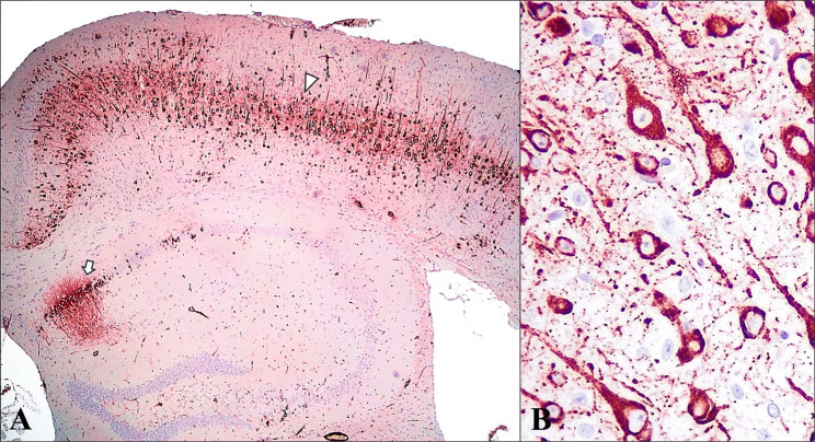
Case presentation: A 4.5-month-old male k18-hACE-2 mouse, part of an experimental study of SARS-CoV-2, displayed a small mass in leptomeningeal area composed by neoplastic round cells. This process is associated with typical acute inflammatory and neurodegenerative lesions according to viral neuroinvasion. Histopathology revealed a well-demarcated tumor composed of lymphoblasts and intermixed with abundant histiocytic-like cells. Immunohistochemistry showed high expression of Iba-1 in histiocytes but negative PAX5, CD3 and IRF-4 labeling. Due to the critical role of PAX-5 in maintaining B-cell function, its reduction or inactivation may favor this loss of identity and differentiation to macrophages, which supports the possibility of a lymphoma undergoing transdifferentiation into a histiocytic/dendritic cells neoplasm. Additionally, SARS-CoV-2 was detected within the tumor histiocytes and adjacent neurons, raising questions about potential interactions between viral infection and tumor development.
Conclusions: While the underlying mechanisms remain uncertain, this finding highlights the need for further investigation into the interplay between SARS-CoV-2 infection and oncogenesis. This case represents the first report of a primary brain histiocytic lymphoproliferative disorder associated with SARS-CoV-2 in k18-hACE2 mouse.


 求助内容:
求助内容: 应助结果提醒方式:


