{"title":"精神分裂症分期、发病部位和病理生理的灰质体积异质性","authors":"Yuchao Jiang, Lena Palaniyappan, Xiao Chang, Jie Zhang, Enpeng Zhou, Xin Yu, Shih-Jen Tsai, Ching-Po Lin, Jingliang Cheng, Yingying Tang, Jijun Wang, Cheng Luo, Dezhong Yao, Long-Biao Cui, Wei Cheng, Jianfeng Feng","doi":"10.1038/s44220-025-00449-9","DOIUrl":null,"url":null,"abstract":"Schizophrenia is characterized with greater variability beyond the mean differences in brain structures. This variability is assumed to be static, reflecting the presence of heterogeneous subgroups, but this assumption and alternative explanations remain untested. Here we examine whether gray matter volume variability decreases in later stages of schizophrenia using magnetic resonance imaging of 1,792 individuals with schizophrenia and 1,523 healthy controls. Compared with healthy controls, greater variability (false-discovery-rate-corrected P < 0.05) was found in 50 regions across the entire patient group. The average variability across all regions was greater in the first-episode than chronic stage (t = 10.8, P = 1.7 × 10⁻⁷). The areas with the largest variability were located at the frontotemporal cortex and thalamus (first-episode), or the hippocampus and caudate (chronic). This study offers novel insights into the diversity of brain alterations in schizophrenia, emphasizing that brain-based heterogeneity is not a static feature; it is more pronounced at the onset of the disorder but reduced over the long term. Schizophrenia exhibits significant variability in brain structures, traditionally viewed as static due to heterogeneous subgroups. Here the authors use magnetic resonance imaging data from 1,792 individuals with schizophrenia to reveal that gray matter volume variability is greater in early stages and decreases over time, highlighting dynamic brain alterations with implications for understanding disease progression.","PeriodicalId":74247,"journal":{"name":"Nature mental health","volume":"3 7","pages":"803-813"},"PeriodicalIF":8.7000,"publicationDate":"2025-06-23","publicationTypes":"Journal Article","fieldsOfStudy":null,"isOpenAccess":false,"openAccessPdf":"","citationCount":"0","resultStr":"{\"title\":\"Gray matter volume heterogeneity by stage, site of origin and pathophysiology in schizophrenia\",\"authors\":\"Yuchao Jiang, Lena Palaniyappan, Xiao Chang, Jie Zhang, Enpeng Zhou, Xin Yu, Shih-Jen Tsai, Ching-Po Lin, Jingliang Cheng, Yingying Tang, Jijun Wang, Cheng Luo, Dezhong Yao, Long-Biao Cui, Wei Cheng, Jianfeng Feng\",\"doi\":\"10.1038/s44220-025-00449-9\",\"DOIUrl\":null,\"url\":null,\"abstract\":\"Schizophrenia is characterized with greater variability beyond the mean differences in brain structures. This variability is assumed to be static, reflecting the presence of heterogeneous subgroups, but this assumption and alternative explanations remain untested. Here we examine whether gray matter volume variability decreases in later stages of schizophrenia using magnetic resonance imaging of 1,792 individuals with schizophrenia and 1,523 healthy controls. Compared with healthy controls, greater variability (false-discovery-rate-corrected P < 0.05) was found in 50 regions across the entire patient group. The average variability across all regions was greater in the first-episode than chronic stage (t = 10.8, P = 1.7 × 10⁻⁷). The areas with the largest variability were located at the frontotemporal cortex and thalamus (first-episode), or the hippocampus and caudate (chronic). This study offers novel insights into the diversity of brain alterations in schizophrenia, emphasizing that brain-based heterogeneity is not a static feature; it is more pronounced at the onset of the disorder but reduced over the long term. Schizophrenia exhibits significant variability in brain structures, traditionally viewed as static due to heterogeneous subgroups. Here the authors use magnetic resonance imaging data from 1,792 individuals with schizophrenia to reveal that gray matter volume variability is greater in early stages and decreases over time, highlighting dynamic brain alterations with implications for understanding disease progression.\",\"PeriodicalId\":74247,\"journal\":{\"name\":\"Nature mental health\",\"volume\":\"3 7\",\"pages\":\"803-813\"},\"PeriodicalIF\":8.7000,\"publicationDate\":\"2025-06-23\",\"publicationTypes\":\"Journal Article\",\"fieldsOfStudy\":null,\"isOpenAccess\":false,\"openAccessPdf\":\"\",\"citationCount\":\"0\",\"resultStr\":null,\"platform\":\"Semanticscholar\",\"paperid\":null,\"PeriodicalName\":\"Nature mental health\",\"FirstCategoryId\":\"1085\",\"ListUrlMain\":\"https://www.nature.com/articles/s44220-025-00449-9\",\"RegionNum\":0,\"RegionCategory\":null,\"ArticlePicture\":[],\"TitleCN\":null,\"AbstractTextCN\":null,\"PMCID\":null,\"EPubDate\":\"\",\"PubModel\":\"\",\"JCR\":\"\",\"JCRName\":\"\",\"Score\":null,\"Total\":0}","platform":"Semanticscholar","paperid":null,"PeriodicalName":"Nature mental health","FirstCategoryId":"1085","ListUrlMain":"https://www.nature.com/articles/s44220-025-00449-9","RegionNum":0,"RegionCategory":null,"ArticlePicture":[],"TitleCN":null,"AbstractTextCN":null,"PMCID":null,"EPubDate":"","PubModel":"","JCR":"","JCRName":"","Score":null,"Total":0}
引用次数: 0
摘要
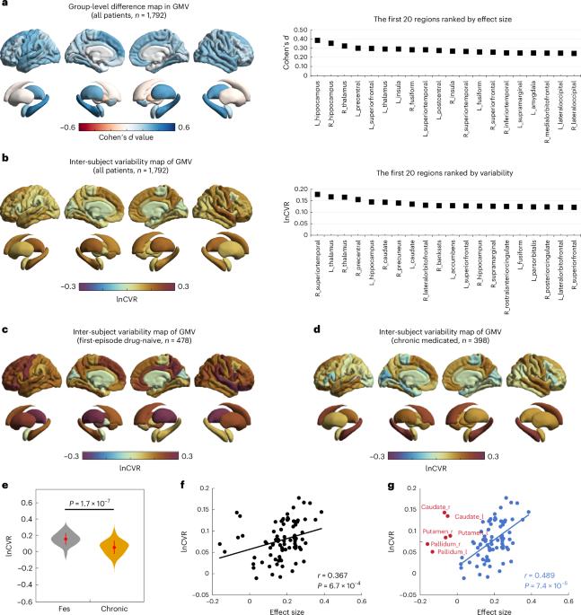
精神分裂症的特点是在大脑结构的平均差异之外有更大的可变性。这种变异性被认为是静态的,反映了异质亚群的存在,但这一假设和其他解释仍未经检验。在这里,我们通过对1792名精神分裂症患者和1523名健康对照者的磁共振成像来研究脑灰质体积变异性是否在精神分裂症晚期减少。与健康对照组相比,在整个患者组的50个地区发现了更大的变异性(经错误发现率校正的P <; 0.05)。所有地区的平均变异性在首发期大于慢性期(t = 10.8, P = 1.7 × 10⁻⁷)。变异最大的区域位于额颞叶皮层和丘脑(首发),或海马和尾状核(慢性)。这项研究为精神分裂症患者大脑改变的多样性提供了新的见解,强调基于大脑的异质性不是一个静态的特征;它在发病时更为明显,但随着时间的推移会逐渐减少。精神分裂症在大脑结构中表现出显著的可变性,传统上认为由于异质性亚群而静态。在这里,作者使用来自1792名精神分裂症患者的磁共振成像数据来揭示灰质体积变异性在早期阶段更大,并随着时间的推移而减少,强调了动态大脑改变对理解疾病进展的影响。
Gray matter volume heterogeneity by stage, site of origin and pathophysiology in schizophrenia
Schizophrenia is characterized with greater variability beyond the mean differences in brain structures. This variability is assumed to be static, reflecting the presence of heterogeneous subgroups, but this assumption and alternative explanations remain untested. Here we examine whether gray matter volume variability decreases in later stages of schizophrenia using magnetic resonance imaging of 1,792 individuals with schizophrenia and 1,523 healthy controls. Compared with healthy controls, greater variability (false-discovery-rate-corrected P < 0.05) was found in 50 regions across the entire patient group. The average variability across all regions was greater in the first-episode than chronic stage (t = 10.8, P = 1.7 × 10⁻⁷). The areas with the largest variability were located at the frontotemporal cortex and thalamus (first-episode), or the hippocampus and caudate (chronic). This study offers novel insights into the diversity of brain alterations in schizophrenia, emphasizing that brain-based heterogeneity is not a static feature; it is more pronounced at the onset of the disorder but reduced over the long term. Schizophrenia exhibits significant variability in brain structures, traditionally viewed as static due to heterogeneous subgroups. Here the authors use magnetic resonance imaging data from 1,792 individuals with schizophrenia to reveal that gray matter volume variability is greater in early stages and decreases over time, highlighting dynamic brain alterations with implications for understanding disease progression.

 求助内容:
求助内容: 应助结果提醒方式:


