利用结构和功能参数的纵向监测对大鼠磁微珠模型进行综合表征
IF 2.7
2区 医学
Q1 OPHTHALMOLOGY
引用次数: 0
摘要
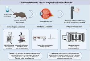
青光眼仍然是世界范围内不可逆失明的主要原因,这推动了对临床前模型的需求,这些模型可以准确地复制人类疾病,并支持开发新的治疗方法。眼内微珠注射是一种广泛应用于高眼压诱导和模拟青光眼病理生理的方法。大多数使用该模型的研究都集中在视网膜神经节细胞(RGC)的损失上,偶尔也会使用视网膜电图(pERG)来评估RGC的功能。在我们的研究中,我们通过结合体内监测的行为、功能和形态评估来扩大模型的表征。此外,我们在该模型中探讨了行为、功能和形态学读数与眼内压(IOP)的关系。采用光谱域光学相干断层扫描(SD-OCT)定量评价视网膜内层(IRL)厚度。记录PERG以评估RGC功能。视动跟踪评估视力(VA)和对比敏感度(CS)。4周后,对大鼠实施安乐死,收集视神经视网膜完整块进行RGC定量和轴突评估。正如预期的那样,我们发现SD-OCT成像证实了IRL的明显变薄。此外,pERG记录显示RGC功能减弱,表现为振幅降低,而视运动测试检测到VA和CS显著下降。IOP与各参数均有显著相关性。磁微珠诱导的高眼压模型成功地模拟了RGCs的结构和功能损伤,早期视觉功能下降与IOP升高有关。结构和功能参数之间的强相关性增强了该模型在探索新的神经保护和再生疗法中的翻译相关性。本文章由计算机程序翻译,如有差异,请以英文原文为准。
Comprehensive characterization of the rat magnetic microbead model using longitudinal monitoring of structural and functional parameters
Glaucoma remains a major cause of irreversible blindness worldwide, driving the need for preclinical models that accurately replicate human disease and support the development of novel treatment methods. Intracameral microbead injection is a widely used method to induce ocular hypertension and mimic the pathophysiology of glaucoma. Most studies using this model focus on retinal ganglion cell (RGC) loss and occasionally pattern electroretinography (pERG) for functional assessment of RGC. In our study, we broaden model characterization by incorporating behavioral, functional, and morphological assessments using in vivo monitoring. In addition, we explored how behavioral, functional, and morphological read-outs correlated with intraocular pressure (IOP) in this model. Spectral-domain optical coherence tomography (SD-OCT) was used for quantitative evaluation of inner retinal layer (IRL) thickness. PERG was recorded to assess RGC function. Optomotor tracking was performed to evaluate visual acuity (VA) and contrast sensitivity (CS). After 4 weeks, the animals were euthanized, and retinal wholemounts with optic nerves were collected for RGC quantification and axon assessment. As expected, we found a significant thinning of IRL as confirmed by SD-OCT imaging. Additionally, pERG recordings revealed diminished RGC function, evidenced by reduced amplitude, whereas optomotor testing detected significant declines in VA and CS. Significant correlations were found between IOP and all parameters. The magnetic microbead-induced ocular hypertension model successfully mimicked structural and functional damage of RGCs, with early visual function declines linked to elevated IOP. Strong correlations between structural and functional parameters enhanced the translational relevance of this model in exploring new neuroprotective and regenerative therapies.
求助全文
通过发布文献求助,成功后即可免费获取论文全文。
去求助
来源期刊

Experimental eye research
医学-眼科学
CiteScore
6.80
自引率
5.90%
发文量
323
审稿时长
66 days
期刊介绍:
The primary goal of Experimental Eye Research is to publish original research papers on all aspects of experimental biology of the eye and ocular tissues that seek to define the mechanisms of normal function and/or disease. Studies of ocular tissues that encompass the disciplines of cell biology, developmental biology, genetics, molecular biology, physiology, biochemistry, biophysics, immunology or microbiology are most welcomed. Manuscripts that are purely clinical or in a surgical area of ophthalmology are not appropriate for submission to Experimental Eye Research and if received will be returned without review.
 求助内容:
求助内容: 应助结果提醒方式:


