基于微计算机断层扫描的Phasmatodea排泄器官的结构组织

IF 2.3 2区 农林科学 Q1 ENTOMOLOGY
Matan Shelomi , Fabian Bäumler , Thies H. Büscher
{"title":"基于微计算机断层扫描的Phasmatodea排泄器官的结构组织","authors":"Matan Shelomi ,&nbsp;Fabian Bäumler ,&nbsp;Thies H. Büscher","doi":"10.1016/j.jinsphys.2025.104889","DOIUrl":null,"url":null,"abstract":"<div><div>Phasmatodea excretory organs differ from other insects’. They consist of two types of Malpighian tubules, excretory and calciferous, and the enigmatic midgut appendages, which are an autapomorphy of this lineage. To gain a better understanding of these three tubules, we used micro-computed tomography to visualize their structural organization in a female <em>Epidares nolimetangere</em>. The midgut appendices were conspicuous, with ducts arising from their ampules penetrating the midgut wall and connecting to the exoperitrophic space. Radio-dense material was observed in the calciferous Malpighian tubules, differentiating them from the excretory Malpighian tubules. While their key role in the development of the biomineralized eggs of Phasmatodea is assumed, the process of how this material is transferred to the eggshells remains unclear. These observations validate previous anatomic and microscopy findings of the Phasmatodea excretory tubules with the description of the structural organization of the excretory organs in a species so far not examined in this regard.</div></div>","PeriodicalId":16189,"journal":{"name":"Journal of insect physiology","volume":"166 ","pages":"Article 104889"},"PeriodicalIF":2.3000,"publicationDate":"2025-09-20","publicationTypes":"Journal Article","fieldsOfStudy":null,"isOpenAccess":false,"openAccessPdf":"","citationCount":"0","resultStr":"{\"title\":\"Structural organization of excretory organs in Phasmatodea based on micro-computed tomography\",\"authors\":\"Matan Shelomi ,&nbsp;Fabian Bäumler ,&nbsp;Thies H. Büscher\",\"doi\":\"10.1016/j.jinsphys.2025.104889\",\"DOIUrl\":null,\"url\":null,\"abstract\":\"<div><div>Phasmatodea excretory organs differ from other insects’. They consist of two types of Malpighian tubules, excretory and calciferous, and the enigmatic midgut appendages, which are an autapomorphy of this lineage. To gain a better understanding of these three tubules, we used micro-computed tomography to visualize their structural organization in a female <em>Epidares nolimetangere</em>. The midgut appendices were conspicuous, with ducts arising from their ampules penetrating the midgut wall and connecting to the exoperitrophic space. Radio-dense material was observed in the calciferous Malpighian tubules, differentiating them from the excretory Malpighian tubules. While their key role in the development of the biomineralized eggs of Phasmatodea is assumed, the process of how this material is transferred to the eggshells remains unclear. These observations validate previous anatomic and microscopy findings of the Phasmatodea excretory tubules with the description of the structural organization of the excretory organs in a species so far not examined in this regard.</div></div>\",\"PeriodicalId\":16189,\"journal\":{\"name\":\"Journal of insect physiology\",\"volume\":\"166 \",\"pages\":\"Article 104889\"},\"PeriodicalIF\":2.3000,\"publicationDate\":\"2025-09-20\",\"publicationTypes\":\"Journal Article\",\"fieldsOfStudy\":null,\"isOpenAccess\":false,\"openAccessPdf\":\"\",\"citationCount\":\"0\",\"resultStr\":null,\"platform\":\"Semanticscholar\",\"paperid\":null,\"PeriodicalName\":\"Journal of insect physiology\",\"FirstCategoryId\":\"97\",\"ListUrlMain\":\"https://www.sciencedirect.com/science/article/pii/S002219102500143X\",\"RegionNum\":2,\"RegionCategory\":\"农林科学\",\"ArticlePicture\":[],\"TitleCN\":null,\"AbstractTextCN\":null,\"PMCID\":null,\"EPubDate\":\"\",\"PubModel\":\"\",\"JCR\":\"Q1\",\"JCRName\":\"ENTOMOLOGY\",\"Score\":null,\"Total\":0}","platform":"Semanticscholar","paperid":null,"PeriodicalName":"Journal of insect physiology","FirstCategoryId":"97","ListUrlMain":"https://www.sciencedirect.com/science/article/pii/S002219102500143X","RegionNum":2,"RegionCategory":"农林科学","ArticlePicture":[],"TitleCN":null,"AbstractTextCN":null,"PMCID":null,"EPubDate":"","PubModel":"","JCR":"Q1","JCRName":"ENTOMOLOGY","Score":null,"Total":0}
引用次数: 0

摘要

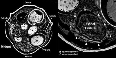
Phasmatodea的排泄器官不同于其他昆虫。它们包括两种类型的马氏小管,排泄小管和钙化小管,以及神秘的中肠附属物,这是该谱系的自异形。为了更好地了解这三个小管,我们使用微型计算机断层扫描来观察雌性小管的结构组织。中肠阑尾很明显,从它们的小体中产生的导管穿过中肠壁并连接到外营养间隙。钙化的马尔比氏小管中可见致密的放射性物质,与排泄的马尔比氏小管相区别。虽然它们在Phasmatodea生物矿化卵的发育中起着关键作用,但这种物质如何转移到蛋壳的过程仍不清楚。这些观察结果证实了先前对Phasmatodea排泄小管的解剖和显微镜观察结果,以及迄今为止尚未在这方面进行研究的物种排泄器官结构组织的描述。
本文章由计算机程序翻译,如有差异,请以英文原文为准。

Structural organization of excretory organs in Phasmatodea based on micro-computed tomography

Structural organization of excretory organs in Phasmatodea based on micro-computed tomography
Phasmatodea excretory organs differ from other insects’. They consist of two types of Malpighian tubules, excretory and calciferous, and the enigmatic midgut appendages, which are an autapomorphy of this lineage. To gain a better understanding of these three tubules, we used micro-computed tomography to visualize their structural organization in a female Epidares nolimetangere. The midgut appendices were conspicuous, with ducts arising from their ampules penetrating the midgut wall and connecting to the exoperitrophic space. Radio-dense material was observed in the calciferous Malpighian tubules, differentiating them from the excretory Malpighian tubules. While their key role in the development of the biomineralized eggs of Phasmatodea is assumed, the process of how this material is transferred to the eggshells remains unclear. These observations validate previous anatomic and microscopy findings of the Phasmatodea excretory tubules with the description of the structural organization of the excretory organs in a species so far not examined in this regard.
求助全文
通过发布文献求助,成功后即可免费获取论文全文。 去求助
来源期刊
Journal of insect physiology
Journal of insect physiology 生物-昆虫学
CiteScore
4.50
自引率
4.50%
发文量
77
审稿时长
57 days
期刊介绍: All aspects of insect physiology are published in this journal which will also accept papers on the physiology of other arthropods, if the referees consider the work to be of general interest. The coverage includes endocrinology (in relation to moulting, reproduction and metabolism), pheromones, neurobiology (cellular, integrative and developmental), physiological pharmacology, nutrition (food selection, digestion and absorption), homeostasis, excretion, reproduction and behaviour. Papers covering functional genomics and molecular approaches to physiological problems will also be included. Communications on structure and applied entomology can be published if the subject matter has an explicit bearing on the physiology of arthropods. Review articles and novel method papers are also welcomed.
×
引用
GB/T 7714-2015
复制
MLA
复制
APA
复制
导出至
BibTeX EndNote RefMan NoteFirst NoteExpress
×
提示
您的信息不完整,为了账户安全,请先补充。
现在去补充
×
提示
您因"违规操作"
具体请查看互助需知
我知道了
×
提示
确定
请完成安全验证×
copy
已复制链接
快去分享给好友吧!
我知道了
右上角分享
点击右上角分享
0
联系我们:info@booksci.cn Book学术提供免费学术资源搜索服务,方便国内外学者检索中英文文献。致力于提供最便捷和优质的服务体验。 Copyright © 2023 布克学术 All rights reserved.
京ICP备2023020795号-1
ghs 京公网安备 11010802042870号
Book学术文献互助
Book学术文献互助群
群 号:604180095
Book学术官方微信