{"title":"主动脉揭密:影像学在主动脉疾病诊断和治疗中的关键作用——综述。","authors":"Jean-Baptiste Ricco, Aurélien Hostalrich, Xavier Chaufour","doi":"10.1093/ehjimp/qyaf108","DOIUrl":null,"url":null,"abstract":"<p><p>Aortic diseases, including aneurysmal and occlusive pathologies of the thoracic and abdominal aorta, represent a significant source of cardiovascular morbidity and mortality. This narrative review explores the role of modern and emerging imaging modalities in the management of aortic disease and highlights the pivotal roles of computed tomography angiography (CTA), magnetic resonance imaging (MRI), and contrast-enhanced ultrasound (CEUS). CTA remains the cornerstone for evaluating aneurysms, dissections, and traumatic injuries, offering high spatial resolution, rapid acquisition, and detailed anatomical assessment. MRI, particularly with advanced sequences such as 4D flow, provides comprehensive multiparametric evaluation without radiation exposure, making it ideal for younger patients and those requiring repeat imaging. Positron emission tomography (PET), especially when integrated with CTA or MRI, enables metabolic characterization of inflammation and infection in aortic walls. Ultrasound, particularly CEUS, remains indispensable in abdominal aortic aneurysm (AAA) screening and post-endovascular aortic aneurysm repair (EVAR) surveillance, especially in patients with renal impairment. Emerging technologies, including hybrid imaging, radiomics, and artificial intelligence (AI) are reshaping the landscape of aortic diagnostics. These innovations enhance detection of subtle imaging features, automate measurements, and may enable prediction of disease progression or complications.</p>","PeriodicalId":94317,"journal":{"name":"European heart journal. Imaging methods and practice","volume":"3 2","pages":"qyaf108"},"PeriodicalIF":0.0000,"publicationDate":"2025-08-13","publicationTypes":"Journal Article","fieldsOfStudy":null,"isOpenAccess":false,"openAccessPdf":"https://www.ncbi.nlm.nih.gov/pmc/articles/PMC12448707/pdf/","citationCount":"0","resultStr":"{\"title\":\"Aorta unveiled: the crucial role of imaging in diagnosing and managing aortic disease-a review.\",\"authors\":\"Jean-Baptiste Ricco, Aurélien Hostalrich, Xavier Chaufour\",\"doi\":\"10.1093/ehjimp/qyaf108\",\"DOIUrl\":null,\"url\":null,\"abstract\":\"<p><p>Aortic diseases, including aneurysmal and occlusive pathologies of the thoracic and abdominal aorta, represent a significant source of cardiovascular morbidity and mortality. This narrative review explores the role of modern and emerging imaging modalities in the management of aortic disease and highlights the pivotal roles of computed tomography angiography (CTA), magnetic resonance imaging (MRI), and contrast-enhanced ultrasound (CEUS). CTA remains the cornerstone for evaluating aneurysms, dissections, and traumatic injuries, offering high spatial resolution, rapid acquisition, and detailed anatomical assessment. MRI, particularly with advanced sequences such as 4D flow, provides comprehensive multiparametric evaluation without radiation exposure, making it ideal for younger patients and those requiring repeat imaging. Positron emission tomography (PET), especially when integrated with CTA or MRI, enables metabolic characterization of inflammation and infection in aortic walls. Ultrasound, particularly CEUS, remains indispensable in abdominal aortic aneurysm (AAA) screening and post-endovascular aortic aneurysm repair (EVAR) surveillance, especially in patients with renal impairment. Emerging technologies, including hybrid imaging, radiomics, and artificial intelligence (AI) are reshaping the landscape of aortic diagnostics. These innovations enhance detection of subtle imaging features, automate measurements, and may enable prediction of disease progression or complications.</p>\",\"PeriodicalId\":94317,\"journal\":{\"name\":\"European heart journal. Imaging methods and practice\",\"volume\":\"3 2\",\"pages\":\"qyaf108\"},\"PeriodicalIF\":0.0000,\"publicationDate\":\"2025-08-13\",\"publicationTypes\":\"Journal Article\",\"fieldsOfStudy\":null,\"isOpenAccess\":false,\"openAccessPdf\":\"https://www.ncbi.nlm.nih.gov/pmc/articles/PMC12448707/pdf/\",\"citationCount\":\"0\",\"resultStr\":null,\"platform\":\"Semanticscholar\",\"paperid\":null,\"PeriodicalName\":\"European heart journal. Imaging methods and practice\",\"FirstCategoryId\":\"1085\",\"ListUrlMain\":\"https://doi.org/10.1093/ehjimp/qyaf108\",\"RegionNum\":0,\"RegionCategory\":null,\"ArticlePicture\":[],\"TitleCN\":null,\"AbstractTextCN\":null,\"PMCID\":null,\"EPubDate\":\"2025/7/1 0:00:00\",\"PubModel\":\"eCollection\",\"JCR\":\"\",\"JCRName\":\"\",\"Score\":null,\"Total\":0}","platform":"Semanticscholar","paperid":null,"PeriodicalName":"European heart journal. Imaging methods and practice","FirstCategoryId":"1085","ListUrlMain":"https://doi.org/10.1093/ehjimp/qyaf108","RegionNum":0,"RegionCategory":null,"ArticlePicture":[],"TitleCN":null,"AbstractTextCN":null,"PMCID":null,"EPubDate":"2025/7/1 0:00:00","PubModel":"eCollection","JCR":"","JCRName":"","Score":null,"Total":0}
Aorta unveiled: the crucial role of imaging in diagnosing and managing aortic disease-a review.
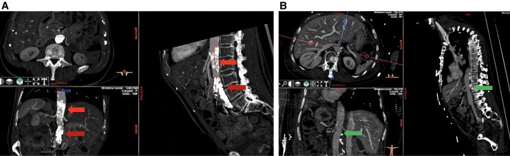
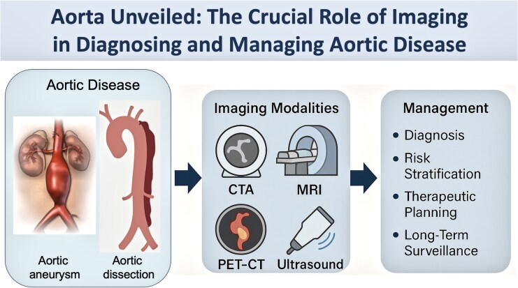
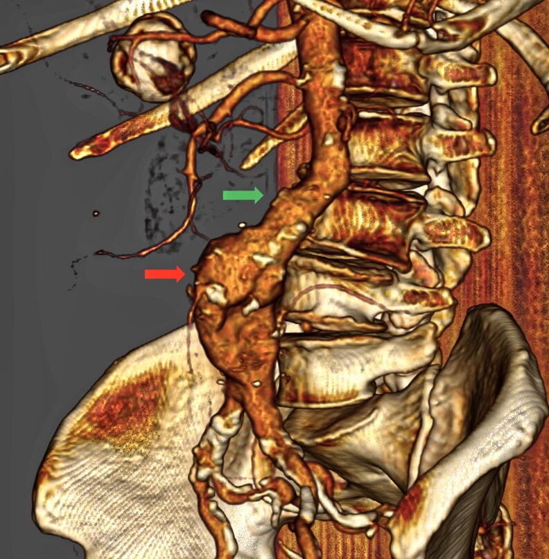
Aortic diseases, including aneurysmal and occlusive pathologies of the thoracic and abdominal aorta, represent a significant source of cardiovascular morbidity and mortality. This narrative review explores the role of modern and emerging imaging modalities in the management of aortic disease and highlights the pivotal roles of computed tomography angiography (CTA), magnetic resonance imaging (MRI), and contrast-enhanced ultrasound (CEUS). CTA remains the cornerstone for evaluating aneurysms, dissections, and traumatic injuries, offering high spatial resolution, rapid acquisition, and detailed anatomical assessment. MRI, particularly with advanced sequences such as 4D flow, provides comprehensive multiparametric evaluation without radiation exposure, making it ideal for younger patients and those requiring repeat imaging. Positron emission tomography (PET), especially when integrated with CTA or MRI, enables metabolic characterization of inflammation and infection in aortic walls. Ultrasound, particularly CEUS, remains indispensable in abdominal aortic aneurysm (AAA) screening and post-endovascular aortic aneurysm repair (EVAR) surveillance, especially in patients with renal impairment. Emerging technologies, including hybrid imaging, radiomics, and artificial intelligence (AI) are reshaping the landscape of aortic diagnostics. These innovations enhance detection of subtle imaging features, automate measurements, and may enable prediction of disease progression or complications.

 求助内容:
求助内容: 应助结果提醒方式:


