{"title":"颈椎MRI显示导致诊断甲状腺功能减退症的低头综合征:1例报告。","authors":"Chiaki Sato, Asako Yamamoto, Megumi Katsumata, Minami Hirasawa, Yuki Hatanaka, Hiroshi Oba","doi":"10.1177/20584601251380870","DOIUrl":null,"url":null,"abstract":"<p><p>Dropped head syndrome, characterized by excessive flexion of the neck, frequently leads to significant impairment in quality of life. Among the various causes of this syndrome, some cases respond effectively to internal medicine. We report a case of a woman in her 70s who presented with dropped head syndrome and was finally diagnosed with hypothyroid myopathy limited to the extensor muscles of the neck. Cervical spine MRI at the initial examination indicated thyroid atrophy, increased subcutaneous fat, and a mild high signal in the right cervical extensor muscles on fat-suppressed T2-weighted images. Blood tests confirmed hypothyroidism. Treatment with levothyroxine improved the symptoms and normalized the blood test results. This case highlights the importance of careful evaluation of the thyroid gland and paravertebral muscles in cervical spine MRI. They can offer diagnostic clues for underlying the important causative role of thyroid disease in dropped head syndrome.</p>","PeriodicalId":72063,"journal":{"name":"Acta radiologica open","volume":"14 9","pages":"20584601251380870"},"PeriodicalIF":1.0000,"publicationDate":"2025-09-18","publicationTypes":"Journal Article","fieldsOfStudy":null,"isOpenAccess":false,"openAccessPdf":"https://www.ncbi.nlm.nih.gov/pmc/articles/PMC12446784/pdf/","citationCount":"0","resultStr":"{\"title\":\"Cervical spine MRI findings leading to diagnosis of hypothyroid myopathy in dropped head syndrome: A case report.\",\"authors\":\"Chiaki Sato, Asako Yamamoto, Megumi Katsumata, Minami Hirasawa, Yuki Hatanaka, Hiroshi Oba\",\"doi\":\"10.1177/20584601251380870\",\"DOIUrl\":null,\"url\":null,\"abstract\":\"<p><p>Dropped head syndrome, characterized by excessive flexion of the neck, frequently leads to significant impairment in quality of life. Among the various causes of this syndrome, some cases respond effectively to internal medicine. We report a case of a woman in her 70s who presented with dropped head syndrome and was finally diagnosed with hypothyroid myopathy limited to the extensor muscles of the neck. Cervical spine MRI at the initial examination indicated thyroid atrophy, increased subcutaneous fat, and a mild high signal in the right cervical extensor muscles on fat-suppressed T2-weighted images. Blood tests confirmed hypothyroidism. Treatment with levothyroxine improved the symptoms and normalized the blood test results. This case highlights the importance of careful evaluation of the thyroid gland and paravertebral muscles in cervical spine MRI. They can offer diagnostic clues for underlying the important causative role of thyroid disease in dropped head syndrome.</p>\",\"PeriodicalId\":72063,\"journal\":{\"name\":\"Acta radiologica open\",\"volume\":\"14 9\",\"pages\":\"20584601251380870\"},\"PeriodicalIF\":1.0000,\"publicationDate\":\"2025-09-18\",\"publicationTypes\":\"Journal Article\",\"fieldsOfStudy\":null,\"isOpenAccess\":false,\"openAccessPdf\":\"https://www.ncbi.nlm.nih.gov/pmc/articles/PMC12446784/pdf/\",\"citationCount\":\"0\",\"resultStr\":null,\"platform\":\"Semanticscholar\",\"paperid\":null,\"PeriodicalName\":\"Acta radiologica open\",\"FirstCategoryId\":\"1085\",\"ListUrlMain\":\"https://doi.org/10.1177/20584601251380870\",\"RegionNum\":0,\"RegionCategory\":null,\"ArticlePicture\":[],\"TitleCN\":null,\"AbstractTextCN\":null,\"PMCID\":null,\"EPubDate\":\"2025/9/1 0:00:00\",\"PubModel\":\"eCollection\",\"JCR\":\"Q4\",\"JCRName\":\"RADIOLOGY, NUCLEAR MEDICINE & MEDICAL IMAGING\",\"Score\":null,\"Total\":0}","platform":"Semanticscholar","paperid":null,"PeriodicalName":"Acta radiologica open","FirstCategoryId":"1085","ListUrlMain":"https://doi.org/10.1177/20584601251380870","RegionNum":0,"RegionCategory":null,"ArticlePicture":[],"TitleCN":null,"AbstractTextCN":null,"PMCID":null,"EPubDate":"2025/9/1 0:00:00","PubModel":"eCollection","JCR":"Q4","JCRName":"RADIOLOGY, NUCLEAR MEDICINE & MEDICAL IMAGING","Score":null,"Total":0}
Cervical spine MRI findings leading to diagnosis of hypothyroid myopathy in dropped head syndrome: A case report.
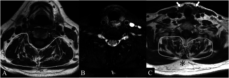
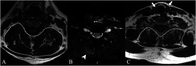
Dropped head syndrome, characterized by excessive flexion of the neck, frequently leads to significant impairment in quality of life. Among the various causes of this syndrome, some cases respond effectively to internal medicine. We report a case of a woman in her 70s who presented with dropped head syndrome and was finally diagnosed with hypothyroid myopathy limited to the extensor muscles of the neck. Cervical spine MRI at the initial examination indicated thyroid atrophy, increased subcutaneous fat, and a mild high signal in the right cervical extensor muscles on fat-suppressed T2-weighted images. Blood tests confirmed hypothyroidism. Treatment with levothyroxine improved the symptoms and normalized the blood test results. This case highlights the importance of careful evaluation of the thyroid gland and paravertebral muscles in cervical spine MRI. They can offer diagnostic clues for underlying the important causative role of thyroid disease in dropped head syndrome.

 求助内容:
求助内容: 应助结果提醒方式:


