Yan Tao, Huanyu Zhao, Sakurako Shimokawa, Masatoshi Fukushima, Kohta Fujiwara, Takahiro Hisai, Kaho Yamamoto, Ayako Okita, Koh-Hei Sonoda, Yusuke Murakami
{"title":"色素性视网膜炎中与黄斑囊样水肿相关的眼部和全身免疫特征。","authors":"Yan Tao, Huanyu Zhao, Sakurako Shimokawa, Masatoshi Fukushima, Kohta Fujiwara, Takahiro Hisai, Kaho Yamamoto, Ayako Okita, Koh-Hei Sonoda, Yusuke Murakami","doi":"10.3389/fopht.2025.1653404","DOIUrl":null,"url":null,"abstract":"<p><strong>Purpose: </strong>We aimed to investigate the local and systemic inflammatory profiles associated with cystoid macular edema (CME) in patients with retinitis pigmentosa (RP).</p><p><strong>Patients and methods: </strong>Paired aqueous humor and serum samples were collected at the time of cataract surgery from 37 eyes of 37 patients with typical RP, including 29 without CME and eight with CME. The concentrations of cytokines and chemokines were determined using a multiplexed immunoassay (Q-Plex). Group comparisons were conducted to assess differences in the inflammatory molecule levels between the RP patients with and without CME. Correlations among the intraocular parameters, the systemic inflammatory molecules, and the CME status were analyzed.</p><p><strong>Results: </strong>Compared to RP patients without CME, those with CME showed significantly increased aqueous levels of interleukin 23 (IL-23) (<i>p</i> = 0.002), I-309 (<i>p</i> = 0.039), and growth-related oncogene alpha (GROα) (<i>p</i> = 0.042). A multiple-factor analysis further supported a potential association between CME formation and an IL-23-related inflammatory network characterized by aqueous IL-23, IL-8, GROα, eotaxin, I-309, serum IL-23, and IFN-γ.</p><p><strong>Conclusion: </strong>These findings suggest that both intraocular and systemic immune activation may play a role in the development of CME in patients with RP. Specifically, IL-23-driven inflammation may be associated with macular fluid accumulation. Further longitudinal studies in larger cohorts are necessary to elucidate these relationships and explore their clinical implications.</p>","PeriodicalId":73096,"journal":{"name":"Frontiers in ophthalmology","volume":"5 ","pages":"1653404"},"PeriodicalIF":0.9000,"publicationDate":"2025-09-05","publicationTypes":"Journal Article","fieldsOfStudy":null,"isOpenAccess":false,"openAccessPdf":"https://www.ncbi.nlm.nih.gov/pmc/articles/PMC12446031/pdf/","citationCount":"0","resultStr":"{\"title\":\"Ocular and systemic immune profiles associated with cystoid macular edema in retinitis pigmentosa.\",\"authors\":\"Yan Tao, Huanyu Zhao, Sakurako Shimokawa, Masatoshi Fukushima, Kohta Fujiwara, Takahiro Hisai, Kaho Yamamoto, Ayako Okita, Koh-Hei Sonoda, Yusuke Murakami\",\"doi\":\"10.3389/fopht.2025.1653404\",\"DOIUrl\":null,\"url\":null,\"abstract\":\"<p><strong>Purpose: </strong>We aimed to investigate the local and systemic inflammatory profiles associated with cystoid macular edema (CME) in patients with retinitis pigmentosa (RP).</p><p><strong>Patients and methods: </strong>Paired aqueous humor and serum samples were collected at the time of cataract surgery from 37 eyes of 37 patients with typical RP, including 29 without CME and eight with CME. The concentrations of cytokines and chemokines were determined using a multiplexed immunoassay (Q-Plex). Group comparisons were conducted to assess differences in the inflammatory molecule levels between the RP patients with and without CME. Correlations among the intraocular parameters, the systemic inflammatory molecules, and the CME status were analyzed.</p><p><strong>Results: </strong>Compared to RP patients without CME, those with CME showed significantly increased aqueous levels of interleukin 23 (IL-23) (<i>p</i> = 0.002), I-309 (<i>p</i> = 0.039), and growth-related oncogene alpha (GROα) (<i>p</i> = 0.042). A multiple-factor analysis further supported a potential association between CME formation and an IL-23-related inflammatory network characterized by aqueous IL-23, IL-8, GROα, eotaxin, I-309, serum IL-23, and IFN-γ.</p><p><strong>Conclusion: </strong>These findings suggest that both intraocular and systemic immune activation may play a role in the development of CME in patients with RP. Specifically, IL-23-driven inflammation may be associated with macular fluid accumulation. Further longitudinal studies in larger cohorts are necessary to elucidate these relationships and explore their clinical implications.</p>\",\"PeriodicalId\":73096,\"journal\":{\"name\":\"Frontiers in ophthalmology\",\"volume\":\"5 \",\"pages\":\"1653404\"},\"PeriodicalIF\":0.9000,\"publicationDate\":\"2025-09-05\",\"publicationTypes\":\"Journal Article\",\"fieldsOfStudy\":null,\"isOpenAccess\":false,\"openAccessPdf\":\"https://www.ncbi.nlm.nih.gov/pmc/articles/PMC12446031/pdf/\",\"citationCount\":\"0\",\"resultStr\":null,\"platform\":\"Semanticscholar\",\"paperid\":null,\"PeriodicalName\":\"Frontiers in ophthalmology\",\"FirstCategoryId\":\"1085\",\"ListUrlMain\":\"https://doi.org/10.3389/fopht.2025.1653404\",\"RegionNum\":0,\"RegionCategory\":null,\"ArticlePicture\":[],\"TitleCN\":null,\"AbstractTextCN\":null,\"PMCID\":null,\"EPubDate\":\"2025/1/1 0:00:00\",\"PubModel\":\"eCollection\",\"JCR\":\"\",\"JCRName\":\"\",\"Score\":null,\"Total\":0}","platform":"Semanticscholar","paperid":null,"PeriodicalName":"Frontiers in ophthalmology","FirstCategoryId":"1085","ListUrlMain":"https://doi.org/10.3389/fopht.2025.1653404","RegionNum":0,"RegionCategory":null,"ArticlePicture":[],"TitleCN":null,"AbstractTextCN":null,"PMCID":null,"EPubDate":"2025/1/1 0:00:00","PubModel":"eCollection","JCR":"","JCRName":"","Score":null,"Total":0}
Ocular and systemic immune profiles associated with cystoid macular edema in retinitis pigmentosa.
Purpose: We aimed to investigate the local and systemic inflammatory profiles associated with cystoid macular edema (CME) in patients with retinitis pigmentosa (RP).
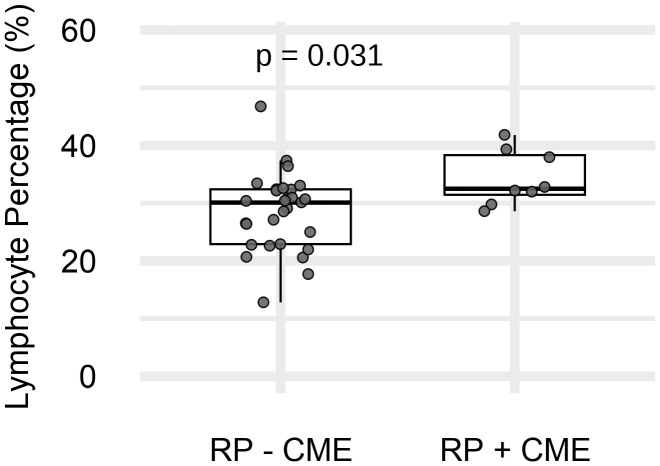
Patients and methods: Paired aqueous humor and serum samples were collected at the time of cataract surgery from 37 eyes of 37 patients with typical RP, including 29 without CME and eight with CME. The concentrations of cytokines and chemokines were determined using a multiplexed immunoassay (Q-Plex). Group comparisons were conducted to assess differences in the inflammatory molecule levels between the RP patients with and without CME. Correlations among the intraocular parameters, the systemic inflammatory molecules, and the CME status were analyzed.
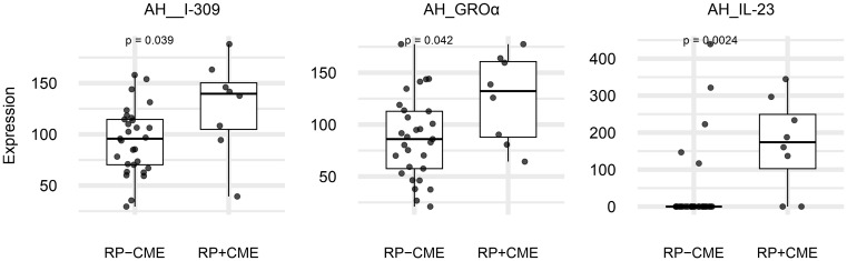
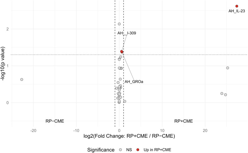
Results: Compared to RP patients without CME, those with CME showed significantly increased aqueous levels of interleukin 23 (IL-23) (p = 0.002), I-309 (p = 0.039), and growth-related oncogene alpha (GROα) (p = 0.042). A multiple-factor analysis further supported a potential association between CME formation and an IL-23-related inflammatory network characterized by aqueous IL-23, IL-8, GROα, eotaxin, I-309, serum IL-23, and IFN-γ.
Conclusion: These findings suggest that both intraocular and systemic immune activation may play a role in the development of CME in patients with RP. Specifically, IL-23-driven inflammation may be associated with macular fluid accumulation. Further longitudinal studies in larger cohorts are necessary to elucidate these relationships and explore their clinical implications.

 求助内容:
求助内容: 应助结果提醒方式:


