Yanqiang Qiao, Yue Qin, Gang Xiao, Lijun Zhang, Jite Shi, Shaohui Ma, Ming Zhang, Wen Gu
{"title":"颈长肌肌腱炎:MRI和临床特征分析与预测疼痛风险模型的发展。","authors":"Yanqiang Qiao, Yue Qin, Gang Xiao, Lijun Zhang, Jite Shi, Shaohui Ma, Ming Zhang, Wen Gu","doi":"10.1155/prm/9211904","DOIUrl":null,"url":null,"abstract":"<p><p><b>Objectives:</b> Longus colli tendinitis (LCT) is a rare, self-limiting disease primarily characterized by neck pain. This study is to investigate and analyze the imaging and clinical features of LCT and to develop a predictive model for pain risk in LCT based on these features. <b>Methods:</b> This study included 35 patients with LCT enrolled between January 2017 and December 2024. Radiological features, laboratory indicators, and clinical profiles were systematically analyzed. We stratified LCT patients into high-risk (<i>n</i> = 20) and low-risk (<i>n</i> = 15) groups based on pain intensity and duration. Nomograms were developed using logistic regression models, with feature selection performed via the least absolute shrinkage and selection operator method. Model performance was evaluated through discrimination (Harrell's C-index) and calibration (calibration plots), with internal validation conducted via bootstrapping. A clinical impact curve was used to assess the model's clinical usefulness. <b>Results:</b> MRI features of LCT included average lesion width of 6.13 mm, length of 64.00 mm, circumference of 134.52 mm, and area of 230.64 mm<sup>2</sup>. Clinically, LCT patients exhibited elevated white blood cell counts, neutrophil counts, hsCRP levels, and IL-6 levels. Feature selection revealed that the lesion area could predict pain risk in LCT patients, which was used to construct a predictive model. The model demonstrated a C-index of 0.93 (95% CI 0.84-0.99). Internal validation confirmed the model's robust performance, with a C-index of 0.93 (95% CI 0.83-0.99). <b>Conclusion:</b> LCT possesses distinct imaging and clinical features. Utilizing these features enables effective prediction of pain risk, thereby assisting clinical decision-making.</p>","PeriodicalId":19913,"journal":{"name":"Pain Research & Management","volume":"2025 ","pages":"9211904"},"PeriodicalIF":3.0000,"publicationDate":"2025-09-11","publicationTypes":"Journal Article","fieldsOfStudy":null,"isOpenAccess":false,"openAccessPdf":"https://www.ncbi.nlm.nih.gov/pmc/articles/PMC12446589/pdf/","citationCount":"0","resultStr":"{\"title\":\"Longus Colli Tendinitis: Analysis of MRI and Clinical Features With Predictive Pain Risk Model Development.\",\"authors\":\"Yanqiang Qiao, Yue Qin, Gang Xiao, Lijun Zhang, Jite Shi, Shaohui Ma, Ming Zhang, Wen Gu\",\"doi\":\"10.1155/prm/9211904\",\"DOIUrl\":null,\"url\":null,\"abstract\":\"<p><p><b>Objectives:</b> Longus colli tendinitis (LCT) is a rare, self-limiting disease primarily characterized by neck pain. This study is to investigate and analyze the imaging and clinical features of LCT and to develop a predictive model for pain risk in LCT based on these features. <b>Methods:</b> This study included 35 patients with LCT enrolled between January 2017 and December 2024. Radiological features, laboratory indicators, and clinical profiles were systematically analyzed. We stratified LCT patients into high-risk (<i>n</i> = 20) and low-risk (<i>n</i> = 15) groups based on pain intensity and duration. Nomograms were developed using logistic regression models, with feature selection performed via the least absolute shrinkage and selection operator method. Model performance was evaluated through discrimination (Harrell's C-index) and calibration (calibration plots), with internal validation conducted via bootstrapping. A clinical impact curve was used to assess the model's clinical usefulness. <b>Results:</b> MRI features of LCT included average lesion width of 6.13 mm, length of 64.00 mm, circumference of 134.52 mm, and area of 230.64 mm<sup>2</sup>. Clinically, LCT patients exhibited elevated white blood cell counts, neutrophil counts, hsCRP levels, and IL-6 levels. Feature selection revealed that the lesion area could predict pain risk in LCT patients, which was used to construct a predictive model. The model demonstrated a C-index of 0.93 (95% CI 0.84-0.99). Internal validation confirmed the model's robust performance, with a C-index of 0.93 (95% CI 0.83-0.99). <b>Conclusion:</b> LCT possesses distinct imaging and clinical features. Utilizing these features enables effective prediction of pain risk, thereby assisting clinical decision-making.</p>\",\"PeriodicalId\":19913,\"journal\":{\"name\":\"Pain Research & Management\",\"volume\":\"2025 \",\"pages\":\"9211904\"},\"PeriodicalIF\":3.0000,\"publicationDate\":\"2025-09-11\",\"publicationTypes\":\"Journal Article\",\"fieldsOfStudy\":null,\"isOpenAccess\":false,\"openAccessPdf\":\"https://www.ncbi.nlm.nih.gov/pmc/articles/PMC12446589/pdf/\",\"citationCount\":\"0\",\"resultStr\":null,\"platform\":\"Semanticscholar\",\"paperid\":null,\"PeriodicalName\":\"Pain Research & Management\",\"FirstCategoryId\":\"3\",\"ListUrlMain\":\"https://doi.org/10.1155/prm/9211904\",\"RegionNum\":3,\"RegionCategory\":\"医学\",\"ArticlePicture\":[],\"TitleCN\":null,\"AbstractTextCN\":null,\"PMCID\":null,\"EPubDate\":\"2025/1/1 0:00:00\",\"PubModel\":\"eCollection\",\"JCR\":\"Q2\",\"JCRName\":\"CLINICAL NEUROLOGY\",\"Score\":null,\"Total\":0}","platform":"Semanticscholar","paperid":null,"PeriodicalName":"Pain Research & Management","FirstCategoryId":"3","ListUrlMain":"https://doi.org/10.1155/prm/9211904","RegionNum":3,"RegionCategory":"医学","ArticlePicture":[],"TitleCN":null,"AbstractTextCN":null,"PMCID":null,"EPubDate":"2025/1/1 0:00:00","PubModel":"eCollection","JCR":"Q2","JCRName":"CLINICAL NEUROLOGY","Score":null,"Total":0}
引用次数: 0
摘要
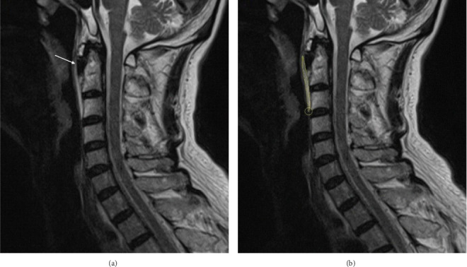
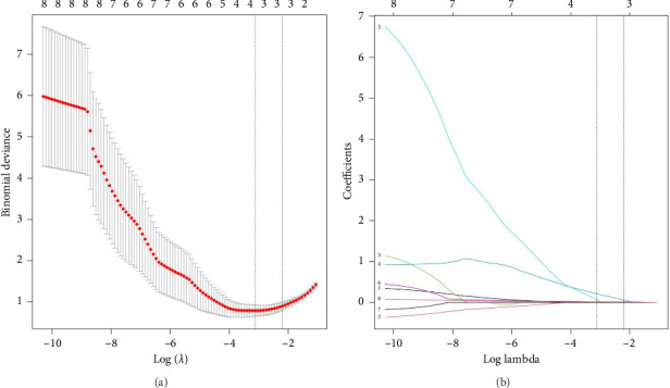
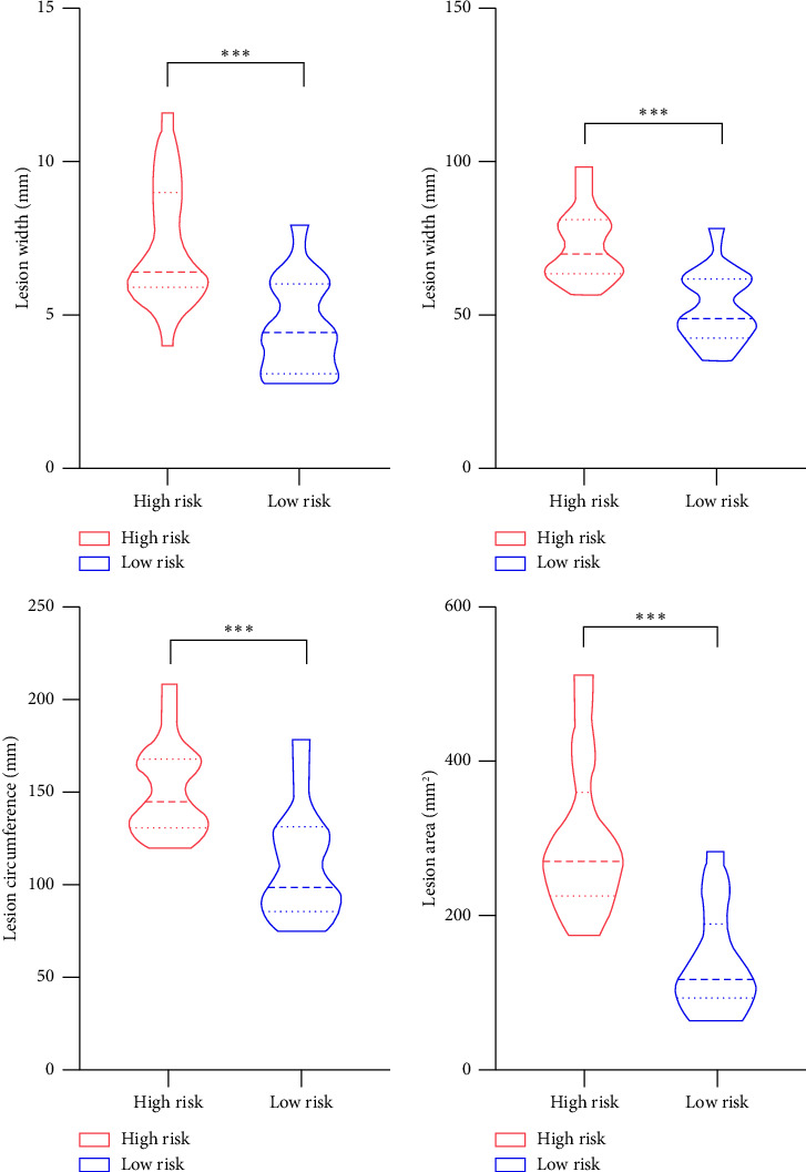
目的:颈长肌腱炎(LCT)是一种罕见的自限性疾病,主要表现为颈部疼痛。本研究旨在调查和分析LCT的影像学和临床特征,并基于这些特征建立LCT疼痛风险的预测模型。方法:本研究纳入了2017年1月至2024年12月期间入选的35例LCT患者。系统分析放射学特征、实验室指标和临床资料。我们根据疼痛强度和持续时间将LCT患者分为高危组(n = 20)和低危组(n = 15)。使用逻辑回归模型开发nomogram,通过最小绝对收缩和选择算子方法进行特征选择。通过判别(Harrell’s C-index)和校准(校准图)对模型性能进行评估,并通过bootstrapping进行内部验证。采用临床影响曲线评估模型的临床有效性。结果:LCT MRI表现为病灶平均宽6.13 mm,长64.00 mm,周长134.52 mm,面积230.64 mm2。临床上,LCT患者表现出白细胞计数、中性粒细胞计数、hsCRP水平和IL-6水平升高。特征选择发现病灶面积可以预测LCT患者的疼痛风险,并以此构建预测模型。该模型的c指数为0.93 (95% CI 0.84-0.99)。内部验证证实了模型的稳健性能,c指数为0.93 (95% CI 0.83-0.99)。结论:LCT具有明显的影像学和临床特点。利用这些特征可以有效地预测疼痛风险,从而帮助临床决策。
Longus Colli Tendinitis: Analysis of MRI and Clinical Features With Predictive Pain Risk Model Development.
Objectives: Longus colli tendinitis (LCT) is a rare, self-limiting disease primarily characterized by neck pain. This study is to investigate and analyze the imaging and clinical features of LCT and to develop a predictive model for pain risk in LCT based on these features. Methods: This study included 35 patients with LCT enrolled between January 2017 and December 2024. Radiological features, laboratory indicators, and clinical profiles were systematically analyzed. We stratified LCT patients into high-risk (n = 20) and low-risk (n = 15) groups based on pain intensity and duration. Nomograms were developed using logistic regression models, with feature selection performed via the least absolute shrinkage and selection operator method. Model performance was evaluated through discrimination (Harrell's C-index) and calibration (calibration plots), with internal validation conducted via bootstrapping. A clinical impact curve was used to assess the model's clinical usefulness. Results: MRI features of LCT included average lesion width of 6.13 mm, length of 64.00 mm, circumference of 134.52 mm, and area of 230.64 mm2. Clinically, LCT patients exhibited elevated white blood cell counts, neutrophil counts, hsCRP levels, and IL-6 levels. Feature selection revealed that the lesion area could predict pain risk in LCT patients, which was used to construct a predictive model. The model demonstrated a C-index of 0.93 (95% CI 0.84-0.99). Internal validation confirmed the model's robust performance, with a C-index of 0.93 (95% CI 0.83-0.99). Conclusion: LCT possesses distinct imaging and clinical features. Utilizing these features enables effective prediction of pain risk, thereby assisting clinical decision-making.
期刊介绍:
Pain Research and Management is a peer-reviewed, Open Access journal that publishes original research articles, review articles, and clinical studies in all areas of pain management.
The most recent Impact Factor for Pain Research and Management is 1.685 according to the 2015 Journal Citation Reports released by Thomson Reuters in 2016.
 求助内容:
求助内容: 应助结果提醒方式:


