{"title":"胃镜下粘膜下剥离时,琥珀红色显像使剥离线更加明显。","authors":"Kohei Funasaka, Ryoji Miyahara, Noriyuki Horiguchi, Hyuga Yamada, Keishi Koyama, Gakushi Komura, Seiya Hagihara, Hijiri Sugiyama, Mizuki Ariga, Mitsuo Nagasaka, Eizaburo Ohno, Teiji Kuzuya, Yoshiki Hirooka","doi":"10.1055/a-2694-7445","DOIUrl":null,"url":null,"abstract":"<p><strong>Background and study aims: </strong>Local injection of a small amount of blue dye into the submucosa can facilitate recognizing the dissection line in endoscopic submucosal dissection (ESD). Amber-red color imaging (ACI), which hardly affects the submucosal blue color, is suitable for the entire ESD. This study aimed to clarify characteristics of ACI during ESD.</p><p><strong>Patients and methods: </strong>Nine endoscopic images were selected during submucosal dissection in four cases of gastric ESD to evaluate endoscopic ACI and white light imaging (WLI). Visibility of the dissection line and the submucosal vessel were evaluated by eight endoscopists using a 5-point Likert scale. The blue submucosal area of each endoscopic image and color signal surrounding the submucosa were compared between ACI and WLI. In addition, the color signals in gradient dilutions of blue solutions were compared in ex vivo experiments.</p><p><strong>Results: </strong>Visibility of the dissection line was better in ACI than in WLI and visibility of the submucosal vessels was slightly better in ACI. The size ratio of the blue area in ACI and WLI (i.e., ACI/WLI) ranged from 0.53 to 0.65, indicating that the blue area in the ACI was narrower. The red signal intensity of the surroundings with respect to the submucosa was greater in ACI than in WLI, which was related to the narrower blue area in ACI. Ex vivo experiments corroborated this observation.</p><p><strong>Conclusions: </strong>ACI highlights the submucosa in blue only where sufficient solution is injected, which facilitates recognition of the dissection line during ESD.</p>","PeriodicalId":11671,"journal":{"name":"Endoscopy International Open","volume":"13 ","pages":"a26947445"},"PeriodicalIF":2.3000,"publicationDate":"2025-09-15","publicationTypes":"Journal Article","fieldsOfStudy":null,"isOpenAccess":false,"openAccessPdf":"https://www.ncbi.nlm.nih.gov/pmc/articles/PMC12445331/pdf/","citationCount":"0","resultStr":"{\"title\":\"Amber-red color imaging makes the dissection line more evident during gastric endoscopic submucosal dissection.\",\"authors\":\"Kohei Funasaka, Ryoji Miyahara, Noriyuki Horiguchi, Hyuga Yamada, Keishi Koyama, Gakushi Komura, Seiya Hagihara, Hijiri Sugiyama, Mizuki Ariga, Mitsuo Nagasaka, Eizaburo Ohno, Teiji Kuzuya, Yoshiki Hirooka\",\"doi\":\"10.1055/a-2694-7445\",\"DOIUrl\":null,\"url\":null,\"abstract\":\"<p><strong>Background and study aims: </strong>Local injection of a small amount of blue dye into the submucosa can facilitate recognizing the dissection line in endoscopic submucosal dissection (ESD). Amber-red color imaging (ACI), which hardly affects the submucosal blue color, is suitable for the entire ESD. This study aimed to clarify characteristics of ACI during ESD.</p><p><strong>Patients and methods: </strong>Nine endoscopic images were selected during submucosal dissection in four cases of gastric ESD to evaluate endoscopic ACI and white light imaging (WLI). Visibility of the dissection line and the submucosal vessel were evaluated by eight endoscopists using a 5-point Likert scale. The blue submucosal area of each endoscopic image and color signal surrounding the submucosa were compared between ACI and WLI. In addition, the color signals in gradient dilutions of blue solutions were compared in ex vivo experiments.</p><p><strong>Results: </strong>Visibility of the dissection line was better in ACI than in WLI and visibility of the submucosal vessels was slightly better in ACI. The size ratio of the blue area in ACI and WLI (i.e., ACI/WLI) ranged from 0.53 to 0.65, indicating that the blue area in the ACI was narrower. The red signal intensity of the surroundings with respect to the submucosa was greater in ACI than in WLI, which was related to the narrower blue area in ACI. Ex vivo experiments corroborated this observation.</p><p><strong>Conclusions: </strong>ACI highlights the submucosa in blue only where sufficient solution is injected, which facilitates recognition of the dissection line during ESD.</p>\",\"PeriodicalId\":11671,\"journal\":{\"name\":\"Endoscopy International Open\",\"volume\":\"13 \",\"pages\":\"a26947445\"},\"PeriodicalIF\":2.3000,\"publicationDate\":\"2025-09-15\",\"publicationTypes\":\"Journal Article\",\"fieldsOfStudy\":null,\"isOpenAccess\":false,\"openAccessPdf\":\"https://www.ncbi.nlm.nih.gov/pmc/articles/PMC12445331/pdf/\",\"citationCount\":\"0\",\"resultStr\":null,\"platform\":\"Semanticscholar\",\"paperid\":null,\"PeriodicalName\":\"Endoscopy International Open\",\"FirstCategoryId\":\"1085\",\"ListUrlMain\":\"https://doi.org/10.1055/a-2694-7445\",\"RegionNum\":0,\"RegionCategory\":null,\"ArticlePicture\":[],\"TitleCN\":null,\"AbstractTextCN\":null,\"PMCID\":null,\"EPubDate\":\"2025/1/1 0:00:00\",\"PubModel\":\"eCollection\",\"JCR\":\"Q3\",\"JCRName\":\"GASTROENTEROLOGY & HEPATOLOGY\",\"Score\":null,\"Total\":0}","platform":"Semanticscholar","paperid":null,"PeriodicalName":"Endoscopy International Open","FirstCategoryId":"1085","ListUrlMain":"https://doi.org/10.1055/a-2694-7445","RegionNum":0,"RegionCategory":null,"ArticlePicture":[],"TitleCN":null,"AbstractTextCN":null,"PMCID":null,"EPubDate":"2025/1/1 0:00:00","PubModel":"eCollection","JCR":"Q3","JCRName":"GASTROENTEROLOGY & HEPATOLOGY","Score":null,"Total":0}
Amber-red color imaging makes the dissection line more evident during gastric endoscopic submucosal dissection.
Background and study aims: Local injection of a small amount of blue dye into the submucosa can facilitate recognizing the dissection line in endoscopic submucosal dissection (ESD). Amber-red color imaging (ACI), which hardly affects the submucosal blue color, is suitable for the entire ESD. This study aimed to clarify characteristics of ACI during ESD.
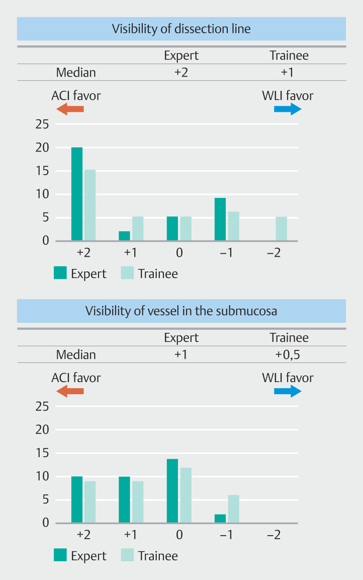
Patients and methods: Nine endoscopic images were selected during submucosal dissection in four cases of gastric ESD to evaluate endoscopic ACI and white light imaging (WLI). Visibility of the dissection line and the submucosal vessel were evaluated by eight endoscopists using a 5-point Likert scale. The blue submucosal area of each endoscopic image and color signal surrounding the submucosa were compared between ACI and WLI. In addition, the color signals in gradient dilutions of blue solutions were compared in ex vivo experiments.
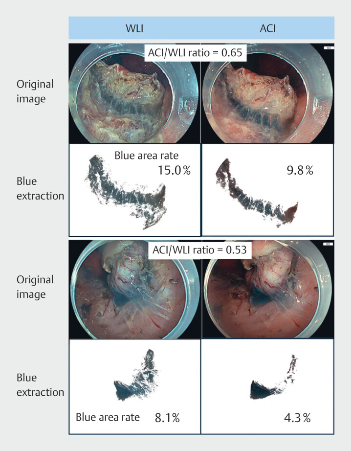
Results: Visibility of the dissection line was better in ACI than in WLI and visibility of the submucosal vessels was slightly better in ACI. The size ratio of the blue area in ACI and WLI (i.e., ACI/WLI) ranged from 0.53 to 0.65, indicating that the blue area in the ACI was narrower. The red signal intensity of the surroundings with respect to the submucosa was greater in ACI than in WLI, which was related to the narrower blue area in ACI. Ex vivo experiments corroborated this observation.
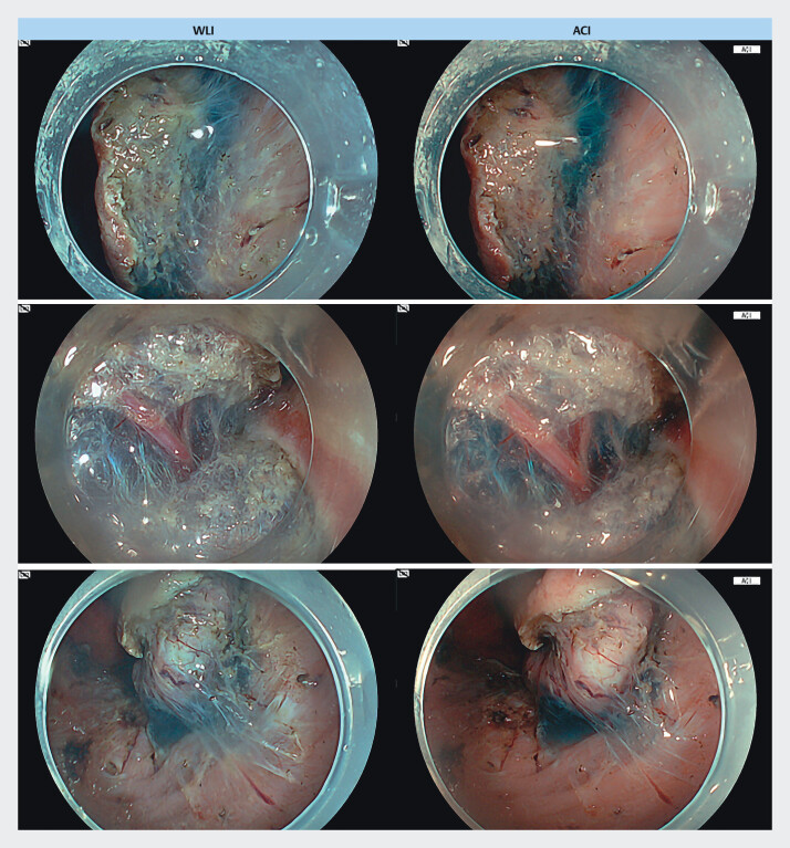
Conclusions: ACI highlights the submucosa in blue only where sufficient solution is injected, which facilitates recognition of the dissection line during ESD.

 求助内容:
求助内容: 应助结果提醒方式:


