仰卧位乳房MRI平面化可视化
IF 2.8
4区 计算机科学
Q2 COMPUTER SCIENCE, SOFTWARE ENGINEERING
引用次数: 0
摘要
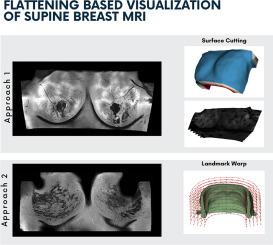
我们提出了两种新的可视化方法,优化了仰卧乳房图像,使乳房组织“变平”,便于检查每个冠状切片内更大的组织区域。乳腺癌是女性中最常见的癌症,早期发现病变对降低死亡率至关重要。仰卧位乳房磁共振成像(MRI)为图像引导干预提供了更好的病灶定位;然而,传统的轴向可视化并不理想,因为组织在胸壁上扩散,导致放射科医生在标准解释时必须滚动浏览许多碎片切片。采用以人为本的设计方法,我们在共同设计和评估扁平化方法的各个阶段都纳入了用户和专家的反馈。我们提出的第一种方法是表面切割方法,它生成偏移曲面并使用尽可能刚性(ARAP)表面网格参数化独立地使其平坦。第二种方法是使用一个基于地标的翘曲,使整个乳房体积一次变平。专家评估表明,表面切割方法提供直观的概述和清晰的血管细节,具有低度量(2-2.5%)和面积(3.7-4.4%)畸变。然而,独立的切片平坦化可以引入层之间的深度扭曲。具有里程碑意义的翘曲提供一致的切片对齐,并支持直接注释和测量,放射科医生因其解剖精度而青睐它。这两种方法都显著减少了需要审查的切片数量,突出了它们节省时间和临床影响的潜力——这是采用仰卧位MRI的重要因素。本文章由计算机程序翻译,如有差异,请以英文原文为准。
Flattening-based visualization of supine breast MRI
We propose two novel visualization methods optimized for supine breast images that “flatten” breast tissue, facilitating examination of larger tissue areas within each coronal slice. Breast cancer is the most frequently diagnosed cancer in women, and early lesion detection is crucial for reducing mortality. Supine breast magnetic resonance imaging (MRI) enables better lesion localization for image-guided interventions; however, traditional axial visualization is suboptimal because the tissue spreads over the chest wall, resulting in numerous fragmented slices that radiologists must scroll through during standard interpretation. Using a human-centered design approach, we incorporated user and expert feedback throughout the co-design and evaluation stages of our flattening methods. Our first proposed method, a surface-cutting approach, generates offset surfaces and flattens them independently using As-Rigid-As-Possible (ARAP) surface mesh parameterization. The second method uses a landmark-based warp to flatten the entire breast volume at once. Expert evaluations revealed that the surface-cutting method provides intuitive overviews and clear vascular detail, with low metric (2–2.5%) and area (3.7–4.4%) distortions. However, independent slice flattening can introduce depth distortions across layers. The landmark warp offers consistent slice alignment and supports direct annotations and measurements, with radiologists favoring it for its anatomical accuracy. Both methods significantly reduced the number of slices needed to review, highlighting their potential for time savings and clinical impact — an essential factor for adopting supine MRI.
求助全文
通过发布文献求助,成功后即可免费获取论文全文。
去求助
来源期刊
Computers & Graphics-Uk
工程技术-计算机:软件工程
CiteScore
5.30
自引率
12.00%
发文量
173
审稿时长
38 days
期刊介绍:
Computers & Graphics is dedicated to disseminate information on research and applications of computer graphics (CG) techniques. The journal encourages articles on:
1. Research and applications of interactive computer graphics. We are particularly interested in novel interaction techniques and applications of CG to problem domains.
2. State-of-the-art papers on late-breaking, cutting-edge research on CG.
3. Information on innovative uses of graphics principles and technologies.
4. Tutorial papers on both teaching CG principles and innovative uses of CG in education.
 求助内容:
求助内容: 应助结果提醒方式:


