{"title":"儿童肾移植术后肾功能及血清细胞因子水平变化的t1制图定量评估","authors":"XueyingWang , Jun Hu","doi":"10.1016/j.cyto.2025.157027","DOIUrl":null,"url":null,"abstract":"<div><div>Background: Postoperative renal function assessment in pediatric kidney transplant recipients faces the challenge of insufficient sensitivity of traditional indicators. T1-mapping, a non-invasive imaging technique, can quantify changes in the microscopic structure of renal tissue. However, its application in the pediatric population and its relationship with serum cytokines remain unclear. This study hypothesized that T1-mapping can quantitatively assess early renal microstructural damage in pediatric kidney transplant recipients and that T1 values correlate with the activation of immune-inflammatory responses (reflected by serum cytokine levels). It aimed to explore the value of T1-mapping in evaluating renal function and its mechanistic association with inflammatory responses.</div><div>Materials and Methods: A total of 31 pediatric kidney transplant recipients (observation group, Obs group) and 31 healthy children (control group, Ctrl group) were enrolled. In the Obs group, T1-mapping was performed at 1, 3, and 6 months post-transplantation to measure T1 values in the renal cortex, medulla, and whole kidney. Serum creatinine (SCr), glomerular filtration rate (GFR), and other renal function indicators were assessed, along with CD4+, CD8+ lymphocyte counts, and levels of cytokines such as interleukin-6 (IL-6) and tumor necrosis factor-alpha (TNF-α). Based on the 6-month postoperative prognosis, participants were divided into the good prognosis group (GPG, <em>n</em> = 20) and poor prognosis group (PPG, <em>n</em> = 11).</div><div>Results: The T1 values of the renal cortex, medulla, and whole kidney in the Obs group were significantly higher than those in the Ctrl group (<em>P</em> < 0.05). Specifically, the cortical T1 value in the PPG was (1820 ± 110) ms, significantly higher than that in the GPG (1650 ± 80) ms (<em>P</em> < 0.05). The SCr in the PPG was (220 ± 35) μmol/L, and the GFR was (22 ± 5) mL/min/1.73m<sup>2</sup>, both significantly worse than the GPG (85 ± 12 μmol/L, 78 ± 10 mL/min/1.73m<sup>2</sup>, <em>P</em> < 0.05). The CD4+/CD8+ ratio in the GPG (1.49 ± 0.21) was higher than that in the PPG (0.87 ± 0.15), while the CD8+ cell count (550 ± 60 × 10<sup>6</sup>/L) in the GPG was lower than that in the PPG (780 ± 75 × 10<sup>6</sup>/L, <em>P</em> < 0.05). Levels of IL-6 (28.8 ± 6.5 pg/mL) and TNF-α (45.5 ± 8.3 pg/mL) in the PPG were significantly higher than those in the GPG (12.5 ± 3.0 pg/mL, 18.2 ± 4.1 pg/mL, <em>P</em> < 0.05).</div><div>Conclusion: T1-mapping technology can quantitatively assess changes in renal function following pediatric kidney transplantation, with increased T1 values closely associated with immune-inflammatory activation and renal function damage. Serum cytokine levels reflect the intensity of the inflammatory response, providing new evidence for postoperative monitoring and intervention.</div></div>","PeriodicalId":297,"journal":{"name":"Cytokine","volume":"196 ","pages":"Article 157027"},"PeriodicalIF":3.7000,"publicationDate":"2025-09-21","publicationTypes":"Journal Article","fieldsOfStudy":null,"isOpenAccess":false,"openAccessPdf":"","citationCount":"0","resultStr":"{\"title\":\"T1-mapping quantitative assessment of renal function and changes in serum cytokine levels after renal transplantation in children\",\"authors\":\"XueyingWang , Jun Hu\",\"doi\":\"10.1016/j.cyto.2025.157027\",\"DOIUrl\":null,\"url\":null,\"abstract\":\"<div><div>Background: Postoperative renal function assessment in pediatric kidney transplant recipients faces the challenge of insufficient sensitivity of traditional indicators. T1-mapping, a non-invasive imaging technique, can quantify changes in the microscopic structure of renal tissue. However, its application in the pediatric population and its relationship with serum cytokines remain unclear. This study hypothesized that T1-mapping can quantitatively assess early renal microstructural damage in pediatric kidney transplant recipients and that T1 values correlate with the activation of immune-inflammatory responses (reflected by serum cytokine levels). It aimed to explore the value of T1-mapping in evaluating renal function and its mechanistic association with inflammatory responses.</div><div>Materials and Methods: A total of 31 pediatric kidney transplant recipients (observation group, Obs group) and 31 healthy children (control group, Ctrl group) were enrolled. In the Obs group, T1-mapping was performed at 1, 3, and 6 months post-transplantation to measure T1 values in the renal cortex, medulla, and whole kidney. Serum creatinine (SCr), glomerular filtration rate (GFR), and other renal function indicators were assessed, along with CD4+, CD8+ lymphocyte counts, and levels of cytokines such as interleukin-6 (IL-6) and tumor necrosis factor-alpha (TNF-α). Based on the 6-month postoperative prognosis, participants were divided into the good prognosis group (GPG, <em>n</em> = 20) and poor prognosis group (PPG, <em>n</em> = 11).</div><div>Results: The T1 values of the renal cortex, medulla, and whole kidney in the Obs group were significantly higher than those in the Ctrl group (<em>P</em> < 0.05). Specifically, the cortical T1 value in the PPG was (1820 ± 110) ms, significantly higher than that in the GPG (1650 ± 80) ms (<em>P</em> < 0.05). The SCr in the PPG was (220 ± 35) μmol/L, and the GFR was (22 ± 5) mL/min/1.73m<sup>2</sup>, both significantly worse than the GPG (85 ± 12 μmol/L, 78 ± 10 mL/min/1.73m<sup>2</sup>, <em>P</em> < 0.05). The CD4+/CD8+ ratio in the GPG (1.49 ± 0.21) was higher than that in the PPG (0.87 ± 0.15), while the CD8+ cell count (550 ± 60 × 10<sup>6</sup>/L) in the GPG was lower than that in the PPG (780 ± 75 × 10<sup>6</sup>/L, <em>P</em> < 0.05). Levels of IL-6 (28.8 ± 6.5 pg/mL) and TNF-α (45.5 ± 8.3 pg/mL) in the PPG were significantly higher than those in the GPG (12.5 ± 3.0 pg/mL, 18.2 ± 4.1 pg/mL, <em>P</em> < 0.05).</div><div>Conclusion: T1-mapping technology can quantitatively assess changes in renal function following pediatric kidney transplantation, with increased T1 values closely associated with immune-inflammatory activation and renal function damage. Serum cytokine levels reflect the intensity of the inflammatory response, providing new evidence for postoperative monitoring and intervention.</div></div>\",\"PeriodicalId\":297,\"journal\":{\"name\":\"Cytokine\",\"volume\":\"196 \",\"pages\":\"Article 157027\"},\"PeriodicalIF\":3.7000,\"publicationDate\":\"2025-09-21\",\"publicationTypes\":\"Journal Article\",\"fieldsOfStudy\":null,\"isOpenAccess\":false,\"openAccessPdf\":\"\",\"citationCount\":\"0\",\"resultStr\":null,\"platform\":\"Semanticscholar\",\"paperid\":null,\"PeriodicalName\":\"Cytokine\",\"FirstCategoryId\":\"3\",\"ListUrlMain\":\"https://www.sciencedirect.com/science/article/pii/S1043466625001747\",\"RegionNum\":3,\"RegionCategory\":\"医学\",\"ArticlePicture\":[],\"TitleCN\":null,\"AbstractTextCN\":null,\"PMCID\":null,\"EPubDate\":\"\",\"PubModel\":\"\",\"JCR\":\"Q2\",\"JCRName\":\"BIOCHEMISTRY & MOLECULAR BIOLOGY\",\"Score\":null,\"Total\":0}","platform":"Semanticscholar","paperid":null,"PeriodicalName":"Cytokine","FirstCategoryId":"3","ListUrlMain":"https://www.sciencedirect.com/science/article/pii/S1043466625001747","RegionNum":3,"RegionCategory":"医学","ArticlePicture":[],"TitleCN":null,"AbstractTextCN":null,"PMCID":null,"EPubDate":"","PubModel":"","JCR":"Q2","JCRName":"BIOCHEMISTRY & MOLECULAR BIOLOGY","Score":null,"Total":0}
引用次数: 0
摘要
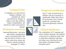
背景:儿童肾移植受者术后肾功能评估面临传统指标敏感性不足的挑战。t1成像是一种非侵入性成像技术,可以量化肾组织微观结构的变化。然而,其在儿童人群中的应用及其与血清细胞因子的关系尚不清楚。本研究假设T1定位可以定量评估儿童肾移植受者早期肾脏微结构损伤,并且T1值与免疫炎症反应的激活相关(通过血清细胞因子水平反映)。旨在探讨t1制图在评估肾功能中的价值及其与炎症反应的机制关联。材料与方法:选取31例儿童肾移植受者(观察组,Obs组)和31例健康儿童(对照组,Ctrl组)作为研究对象。在Obs组,在移植后1、3和6个月进行T1制图,测量肾皮质、髓质和全肾的T1值。评估血清肌酐(SCr)、肾小球滤过率(GFR)和其他肾功能指标,以及CD4+、CD8+淋巴细胞计数,以及白细胞介素-6 (IL-6)和肿瘤坏死因子-α (TNF-α)等细胞因子水平。根据术后6个月预后分为预后良好组(GPG, n = 20)和预后不良组(PPG, n = 11)。结果:Obs组肾皮质、髓质、全肾T1值均显著高于对照组(P < 0.05)。其中,PPG组皮层T1值为(1820±110)ms,显著高于GPG组(1650±80)ms (P < 0.05)。PPG的SCr为(220±35)μmol/L, GFR为(22±5)mL/min/1.73m2,均显著低于GPG(85±12 μmol/L, 78±10 mL/min/1.73m2, P < 0.05)。GPG中CD4+/CD8+比值(1.49±0.21)高于PPG(0.87±0.15),而CD8+细胞计数(550±60 × 106/L)低于PPG(780±75 × 106/L, P < 0.05)。PPG组IL-6(28.8±6.5 pg/mL)、TNF-α(45.5±8.3 pg/mL)水平显著高于GPG组(12.5±3.0 pg/mL、18.2±4.1 pg/mL, P < 0.05)。结论:T1制图技术可以定量评估儿童肾移植术后肾功能的变化,T1值升高与免疫炎症激活和肾功能损害密切相关。血清细胞因子水平反映炎症反应的强度,为术后监测和干预提供新的依据。
T1-mapping quantitative assessment of renal function and changes in serum cytokine levels after renal transplantation in children
Background: Postoperative renal function assessment in pediatric kidney transplant recipients faces the challenge of insufficient sensitivity of traditional indicators. T1-mapping, a non-invasive imaging technique, can quantify changes in the microscopic structure of renal tissue. However, its application in the pediatric population and its relationship with serum cytokines remain unclear. This study hypothesized that T1-mapping can quantitatively assess early renal microstructural damage in pediatric kidney transplant recipients and that T1 values correlate with the activation of immune-inflammatory responses (reflected by serum cytokine levels). It aimed to explore the value of T1-mapping in evaluating renal function and its mechanistic association with inflammatory responses.
Materials and Methods: A total of 31 pediatric kidney transplant recipients (observation group, Obs group) and 31 healthy children (control group, Ctrl group) were enrolled. In the Obs group, T1-mapping was performed at 1, 3, and 6 months post-transplantation to measure T1 values in the renal cortex, medulla, and whole kidney. Serum creatinine (SCr), glomerular filtration rate (GFR), and other renal function indicators were assessed, along with CD4+, CD8+ lymphocyte counts, and levels of cytokines such as interleukin-6 (IL-6) and tumor necrosis factor-alpha (TNF-α). Based on the 6-month postoperative prognosis, participants were divided into the good prognosis group (GPG, n = 20) and poor prognosis group (PPG, n = 11).
Results: The T1 values of the renal cortex, medulla, and whole kidney in the Obs group were significantly higher than those in the Ctrl group (P < 0.05). Specifically, the cortical T1 value in the PPG was (1820 ± 110) ms, significantly higher than that in the GPG (1650 ± 80) ms (P < 0.05). The SCr in the PPG was (220 ± 35) μmol/L, and the GFR was (22 ± 5) mL/min/1.73m2, both significantly worse than the GPG (85 ± 12 μmol/L, 78 ± 10 mL/min/1.73m2, P < 0.05). The CD4+/CD8+ ratio in the GPG (1.49 ± 0.21) was higher than that in the PPG (0.87 ± 0.15), while the CD8+ cell count (550 ± 60 × 106/L) in the GPG was lower than that in the PPG (780 ± 75 × 106/L, P < 0.05). Levels of IL-6 (28.8 ± 6.5 pg/mL) and TNF-α (45.5 ± 8.3 pg/mL) in the PPG were significantly higher than those in the GPG (12.5 ± 3.0 pg/mL, 18.2 ± 4.1 pg/mL, P < 0.05).
Conclusion: T1-mapping technology can quantitatively assess changes in renal function following pediatric kidney transplantation, with increased T1 values closely associated with immune-inflammatory activation and renal function damage. Serum cytokine levels reflect the intensity of the inflammatory response, providing new evidence for postoperative monitoring and intervention.
期刊介绍:
The journal Cytokine has an open access mirror journal Cytokine: X, sharing the same aims and scope, editorial team, submission system and rigorous peer review.
* Devoted exclusively to the study of the molecular biology, genetics, biochemistry, immunology, genome-wide association studies, pathobiology, diagnostic and clinical applications of all known interleukins, hematopoietic factors, growth factors, cytotoxins, interferons, new cytokines, and chemokines, Cytokine provides comprehensive coverage of cytokines and their mechanisms of actions, 12 times a year by publishing original high quality refereed scientific papers from prominent investigators in both the academic and industrial sectors.
We will publish 3 major types of manuscripts:
1) Original manuscripts describing research results.
2) Basic and clinical reviews describing cytokine actions and regulation.
3) Short commentaries/perspectives on recently published aspects of cytokines, pathogenesis and clinical results.