Wenjing Wang , Ye He , Daheng Liang, Liuhua Zhou, Tongtong Yang, Lingfeng Gu, Chong Du, Sibo Wang, Hao Wang, Liansheng Wang, Qiming Wang
{"title":"成纤维细胞分泌的ADAMTSL2通过激活LRP6/β-catenin信号通路促进心肌梗死后的心脏修复。","authors":"Wenjing Wang , Ye He , Daheng Liang, Liuhua Zhou, Tongtong Yang, Lingfeng Gu, Chong Du, Sibo Wang, Hao Wang, Liansheng Wang, Qiming Wang","doi":"10.1016/j.cellsig.2025.112144","DOIUrl":null,"url":null,"abstract":"<div><div>When coronary reperfusion is delayed in myocardial infarction (MI), persistent ischemic injury induces progressive loss of cardiomyocytes, ultimately resulting in pathological ventricular remodeling and heart failure. Secreted proteins may play a critical role in modulating cardiomyocyte death following MI, offering potential therapeutic targets. This study elucidated the biological function of fibroblast-secreted ADAMTSL2 in cardiomyocyte apoptosis post-MI. Elevated ADAMTSL2 levels were detected by ELISA in the serum of acute myocardial infarction patients and by immunoblotting in the infarcted myocardium of mice. Hypoxia treatment significantly upregulated ADAMTSL2 expression in neonatal rat cardiac fibroblasts (NRCFs), whereas no such hypoxic response was observed in neonatal rat cardiomyocytes (NRCMs). In vitro, overexpression of ADAMTSL2 in NRCFs attenuated oxygen-glucose deprivation (OGD)-induced apoptosis in NRCMs, whereas knockdown of ADAMTSL2 in NRCFs exacerbated cardiomyocyte apoptosis. In vivo, fibroblast-targeted overexpression of ADAMTSL2 by adenovirus 5 significantly reduced cardiomyocyte apoptosis and ameliorated adverse left ventricular remodeling post-MI. Conversely, ADAMTSL2 knockdown exacerbated infarct size and accelerated pathological remodeling. Mechanistically, ADAMTSL2 overexpression increased the expression of β-catenin in cardiomyocytes. Co-immunoprecipitation (Co-IP) assay showed that ADAMTSL2 could directly bind to LRP6 and promote its phosphorylation, leading to β-catenin stabilization and subsequent nuclear translocation. In summary, our study indicates that ADAMTSL2 protects against cardiomyocyte apoptosis and promotes cardiac repair by activating LRP6/β-catenin signaling.</div></div>","PeriodicalId":9902,"journal":{"name":"Cellular signalling","volume":"136 ","pages":"Article 112144"},"PeriodicalIF":3.7000,"publicationDate":"2025-09-18","publicationTypes":"Journal Article","fieldsOfStudy":null,"isOpenAccess":false,"openAccessPdf":"","citationCount":"0","resultStr":"{\"title\":\"Fibroblast-secreted ADAMTSL2 promotes cardiac repair after myocardial infarction by activating LRP6/β-catenin signaling\",\"authors\":\"Wenjing Wang , Ye He , Daheng Liang, Liuhua Zhou, Tongtong Yang, Lingfeng Gu, Chong Du, Sibo Wang, Hao Wang, Liansheng Wang, Qiming Wang\",\"doi\":\"10.1016/j.cellsig.2025.112144\",\"DOIUrl\":null,\"url\":null,\"abstract\":\"<div><div>When coronary reperfusion is delayed in myocardial infarction (MI), persistent ischemic injury induces progressive loss of cardiomyocytes, ultimately resulting in pathological ventricular remodeling and heart failure. Secreted proteins may play a critical role in modulating cardiomyocyte death following MI, offering potential therapeutic targets. This study elucidated the biological function of fibroblast-secreted ADAMTSL2 in cardiomyocyte apoptosis post-MI. Elevated ADAMTSL2 levels were detected by ELISA in the serum of acute myocardial infarction patients and by immunoblotting in the infarcted myocardium of mice. Hypoxia treatment significantly upregulated ADAMTSL2 expression in neonatal rat cardiac fibroblasts (NRCFs), whereas no such hypoxic response was observed in neonatal rat cardiomyocytes (NRCMs). In vitro, overexpression of ADAMTSL2 in NRCFs attenuated oxygen-glucose deprivation (OGD)-induced apoptosis in NRCMs, whereas knockdown of ADAMTSL2 in NRCFs exacerbated cardiomyocyte apoptosis. In vivo, fibroblast-targeted overexpression of ADAMTSL2 by adenovirus 5 significantly reduced cardiomyocyte apoptosis and ameliorated adverse left ventricular remodeling post-MI. Conversely, ADAMTSL2 knockdown exacerbated infarct size and accelerated pathological remodeling. Mechanistically, ADAMTSL2 overexpression increased the expression of β-catenin in cardiomyocytes. Co-immunoprecipitation (Co-IP) assay showed that ADAMTSL2 could directly bind to LRP6 and promote its phosphorylation, leading to β-catenin stabilization and subsequent nuclear translocation. In summary, our study indicates that ADAMTSL2 protects against cardiomyocyte apoptosis and promotes cardiac repair by activating LRP6/β-catenin signaling.</div></div>\",\"PeriodicalId\":9902,\"journal\":{\"name\":\"Cellular signalling\",\"volume\":\"136 \",\"pages\":\"Article 112144\"},\"PeriodicalIF\":3.7000,\"publicationDate\":\"2025-09-18\",\"publicationTypes\":\"Journal Article\",\"fieldsOfStudy\":null,\"isOpenAccess\":false,\"openAccessPdf\":\"\",\"citationCount\":\"0\",\"resultStr\":null,\"platform\":\"Semanticscholar\",\"paperid\":null,\"PeriodicalName\":\"Cellular signalling\",\"FirstCategoryId\":\"99\",\"ListUrlMain\":\"https://www.sciencedirect.com/science/article/pii/S0898656825005595\",\"RegionNum\":2,\"RegionCategory\":\"生物学\",\"ArticlePicture\":[],\"TitleCN\":null,\"AbstractTextCN\":null,\"PMCID\":null,\"EPubDate\":\"\",\"PubModel\":\"\",\"JCR\":\"Q2\",\"JCRName\":\"CELL BIOLOGY\",\"Score\":null,\"Total\":0}","platform":"Semanticscholar","paperid":null,"PeriodicalName":"Cellular signalling","FirstCategoryId":"99","ListUrlMain":"https://www.sciencedirect.com/science/article/pii/S0898656825005595","RegionNum":2,"RegionCategory":"生物学","ArticlePicture":[],"TitleCN":null,"AbstractTextCN":null,"PMCID":null,"EPubDate":"","PubModel":"","JCR":"Q2","JCRName":"CELL BIOLOGY","Score":null,"Total":0}
Fibroblast-secreted ADAMTSL2 promotes cardiac repair after myocardial infarction by activating LRP6/β-catenin signaling
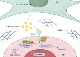
When coronary reperfusion is delayed in myocardial infarction (MI), persistent ischemic injury induces progressive loss of cardiomyocytes, ultimately resulting in pathological ventricular remodeling and heart failure. Secreted proteins may play a critical role in modulating cardiomyocyte death following MI, offering potential therapeutic targets. This study elucidated the biological function of fibroblast-secreted ADAMTSL2 in cardiomyocyte apoptosis post-MI. Elevated ADAMTSL2 levels were detected by ELISA in the serum of acute myocardial infarction patients and by immunoblotting in the infarcted myocardium of mice. Hypoxia treatment significantly upregulated ADAMTSL2 expression in neonatal rat cardiac fibroblasts (NRCFs), whereas no such hypoxic response was observed in neonatal rat cardiomyocytes (NRCMs). In vitro, overexpression of ADAMTSL2 in NRCFs attenuated oxygen-glucose deprivation (OGD)-induced apoptosis in NRCMs, whereas knockdown of ADAMTSL2 in NRCFs exacerbated cardiomyocyte apoptosis. In vivo, fibroblast-targeted overexpression of ADAMTSL2 by adenovirus 5 significantly reduced cardiomyocyte apoptosis and ameliorated adverse left ventricular remodeling post-MI. Conversely, ADAMTSL2 knockdown exacerbated infarct size and accelerated pathological remodeling. Mechanistically, ADAMTSL2 overexpression increased the expression of β-catenin in cardiomyocytes. Co-immunoprecipitation (Co-IP) assay showed that ADAMTSL2 could directly bind to LRP6 and promote its phosphorylation, leading to β-catenin stabilization and subsequent nuclear translocation. In summary, our study indicates that ADAMTSL2 protects against cardiomyocyte apoptosis and promotes cardiac repair by activating LRP6/β-catenin signaling.
期刊介绍:
Cellular Signalling publishes original research describing fundamental and clinical findings on the mechanisms, actions and structural components of cellular signalling systems in vitro and in vivo.
Cellular Signalling aims at full length research papers defining signalling systems ranging from microorganisms to cells, tissues and higher organisms.