磁共振成像在评估活体肝移植供者资格中的作用:现状和未来方向
IF 3.2
Q2 GASTROENTEROLOGY & HEPATOLOGY
Journal of Clinical and Experimental Hepatology
Pub Date : 2025-09-01
DOI:10.1016/j.jceh.2025.103182
引用次数: 0
摘要
对比增强计算机断层扫描(CECT)评估潜在的活体肝移植(LDLT)供体是供体资格测试的一个既定组成部分。通常进行非对比磁共振成像(MRI)的目的是评估胆道解剖和肝脏脂肪分数。虽然少数供者被认为不适合在CECT后进行LDLT,主要是由于中度肝脂肪变性或肝残余不足,但其他肝脏或肝外异常也可能排除捐赠。血管解剖学知识对于外科医生的指导是必不可少的,但随着手术技术和专业知识的发展,它很少成为供体排斥的原因。非对比MRI可用于全面筛查合格的LDLT供体,甚至在CECT评估之前,因为它提供了详细的肝脏和肝外腹部评估以及体积估计,而无需任何额外支出。这种做法不仅有助于避免不合适供体的过度暴露于CT辐射和碘化造影剂,而且还可以利用基于mri的可靠肝脏脂肪估计,为具有临界高脂肪含量的边缘供体的移植前减重提供指导。本文章由计算机程序翻译,如有差异,请以英文原文为准。
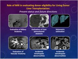
Role of Magnetic Resonance Imaging in Evaluating Donor Eligibility for Living Donor Liver Transplantation: Present Status and Future Directions
Contrast-enhanced computed tomography (CECT) evaluation of a potential living donor liver transplantation (LDLT) donor is an established component of donor eligibility tests. Usually noncontrast magnetic resonance imaging (MRI) is performed with the aim of assessing biliary anatomy and liver fat fraction. While a few donors are considered ineligible for LDLT after CECT, primarily due to moderate liver steatosis or inadequate liver remnant, other hepatic or extrahepatic abnormalities may also preclude donation. Knowledge regarding vascular anatomy is essential to provide a roadmap to the surgeon but is seldom a reason for donor rejection with the developments in surgical technique and expertise.
Noncontrast MRI can be utilized to comprehensively screen eligible LDLT donors, even before CECT evaluation, as it provides a detailed hepatic and extrahepatic abdominal evaluation along with volumetric estimation without any extra expenditure. This practice not only helps to avoid undue exposure to CT radiation and iodinated contrast in unsuitable donors but also provides guidance for pretransplant modifications in terms of weight reduction in marginal donors with borderline high-fat content by taking advantage of the robust MRI-based liver fat estimation.
求助全文
通过发布文献求助,成功后即可免费获取论文全文。
去求助
来源期刊
Journal of Clinical and Experimental Hepatology
GASTROENTEROLOGY & HEPATOLOGY-
CiteScore
4.90
自引率
16.70%
发文量
537
审稿时长
64 days
 求助内容:
求助内容: 应助结果提醒方式:


