Hartmut Häntze, Lina Xu, Maximilian Nikolas Rattunde, Leonhard Donle, Felix J Dorfner, Alessa Hering, Jawed Nawabi, Lisa C Adams, Keno K Bressem
{"title":"基于反演预处理的CT模型自适应MRI注释。","authors":"Hartmut Häntze, Lina Xu, Maximilian Nikolas Rattunde, Leonhard Donle, Felix J Dorfner, Alessa Hering, Jawed Nawabi, Lisa C Adams, Keno K Bressem","doi":"10.1186/s41747-025-00626-6","DOIUrl":null,"url":null,"abstract":"<p><strong>Background: </strong>Annotating new classes in MRI images is time-consuming. Refining presegmented structures can accelerate this process. Many target classes lacking in MRI are supported by computed tomography (CT) models, but translating MRI to synthetic CT images is challenging. We demonstrate that CT segmentation models can create accurate MRI presegmentations, with or without image inversion.</p><p><strong>Materials and methods: </strong>We retrospectively investigated the performance of two CT-trained models on MRI images: a general multiclass model (TotalSegmentator); and a specialized renal tumor model trained in-house. Both models were applied to 100 T1-weighted (T1w) and 100 T2-weighted fat-saturated (T2wfs) MRI sequences from 100 patients (50 male). Segmentation quality was evaluated on both raw and intensity-inverted sequences using Dice similarity coefficients (DSC), with reference annotations comprising manual kidney tumor annotations and automatically generated segmentations for 24 abdominal structures.</p><p><strong>Results: </strong>Segmentation quality varied by MRI sequence and anatomical structure. Both models accurately segmented kidneys in T2wfs sequences without preprocessing (TotalSegmentator DSC 0.60), but TotalSegmentator failed to segment blood vessels and muscles. In T1w sequences, intensity inversion significantly improved TotalSegmentator performance, increasing the mean DSC across 24 structures from 0.04 to 0.56 (p < 0.001). Kidney tumor segmentation demonstrated poor performance in T2wfs sequences regardless of preprocessing. In T1w sequences, inversion improved tumor segmentation DSC from 0.04 to 0.42 (p < 0.001).</p><p><strong>Conclusion: </strong>CT-trained models can generalize to MRI when supported by image augmentation. Inversion preprocessing enabled segmentation of renal cell carcinoma in T1w MRI using a CT-trained model. CT models might be transferable to the MRI domain.</p><p><strong>Relevance statement: </strong>CT-trained artificial intelligence models can be adapted for MRI segmentation using simple preprocessing, potentially reducing manual annotation efforts and accelerating the development of AI-assisted tools for MRI analysis in research and future clinical practice.</p><p><strong>Key points: </strong>CT segmentation models can create presegmentations for many structures in MRI scans. T1w MRI scans require an additional inversion step before segmenting with a CT model. Results were consistent for a large multiclass model (i.e., TotalSegmentator) and a smaller model for renal cell carcinoma.</p>","PeriodicalId":36926,"journal":{"name":"European Radiology Experimental","volume":"9 1","pages":"93"},"PeriodicalIF":3.6000,"publicationDate":"2025-09-19","publicationTypes":"Journal Article","fieldsOfStudy":null,"isOpenAccess":false,"openAccessPdf":"https://www.ncbi.nlm.nih.gov/pmc/articles/PMC12449280/pdf/","citationCount":"0","resultStr":"{\"title\":\"MRI annotation using an inversion-based preprocessing for CT model adaptation.\",\"authors\":\"Hartmut Häntze, Lina Xu, Maximilian Nikolas Rattunde, Leonhard Donle, Felix J Dorfner, Alessa Hering, Jawed Nawabi, Lisa C Adams, Keno K Bressem\",\"doi\":\"10.1186/s41747-025-00626-6\",\"DOIUrl\":null,\"url\":null,\"abstract\":\"<p><strong>Background: </strong>Annotating new classes in MRI images is time-consuming. Refining presegmented structures can accelerate this process. Many target classes lacking in MRI are supported by computed tomography (CT) models, but translating MRI to synthetic CT images is challenging. We demonstrate that CT segmentation models can create accurate MRI presegmentations, with or without image inversion.</p><p><strong>Materials and methods: </strong>We retrospectively investigated the performance of two CT-trained models on MRI images: a general multiclass model (TotalSegmentator); and a specialized renal tumor model trained in-house. Both models were applied to 100 T1-weighted (T1w) and 100 T2-weighted fat-saturated (T2wfs) MRI sequences from 100 patients (50 male). Segmentation quality was evaluated on both raw and intensity-inverted sequences using Dice similarity coefficients (DSC), with reference annotations comprising manual kidney tumor annotations and automatically generated segmentations for 24 abdominal structures.</p><p><strong>Results: </strong>Segmentation quality varied by MRI sequence and anatomical structure. Both models accurately segmented kidneys in T2wfs sequences without preprocessing (TotalSegmentator DSC 0.60), but TotalSegmentator failed to segment blood vessels and muscles. In T1w sequences, intensity inversion significantly improved TotalSegmentator performance, increasing the mean DSC across 24 structures from 0.04 to 0.56 (p < 0.001). Kidney tumor segmentation demonstrated poor performance in T2wfs sequences regardless of preprocessing. In T1w sequences, inversion improved tumor segmentation DSC from 0.04 to 0.42 (p < 0.001).</p><p><strong>Conclusion: </strong>CT-trained models can generalize to MRI when supported by image augmentation. Inversion preprocessing enabled segmentation of renal cell carcinoma in T1w MRI using a CT-trained model. CT models might be transferable to the MRI domain.</p><p><strong>Relevance statement: </strong>CT-trained artificial intelligence models can be adapted for MRI segmentation using simple preprocessing, potentially reducing manual annotation efforts and accelerating the development of AI-assisted tools for MRI analysis in research and future clinical practice.</p><p><strong>Key points: </strong>CT segmentation models can create presegmentations for many structures in MRI scans. T1w MRI scans require an additional inversion step before segmenting with a CT model. Results were consistent for a large multiclass model (i.e., TotalSegmentator) and a smaller model for renal cell carcinoma.</p>\",\"PeriodicalId\":36926,\"journal\":{\"name\":\"European Radiology Experimental\",\"volume\":\"9 1\",\"pages\":\"93\"},\"PeriodicalIF\":3.6000,\"publicationDate\":\"2025-09-19\",\"publicationTypes\":\"Journal Article\",\"fieldsOfStudy\":null,\"isOpenAccess\":false,\"openAccessPdf\":\"https://www.ncbi.nlm.nih.gov/pmc/articles/PMC12449280/pdf/\",\"citationCount\":\"0\",\"resultStr\":null,\"platform\":\"Semanticscholar\",\"paperid\":null,\"PeriodicalName\":\"European Radiology Experimental\",\"FirstCategoryId\":\"1085\",\"ListUrlMain\":\"https://doi.org/10.1186/s41747-025-00626-6\",\"RegionNum\":0,\"RegionCategory\":null,\"ArticlePicture\":[],\"TitleCN\":null,\"AbstractTextCN\":null,\"PMCID\":null,\"EPubDate\":\"\",\"PubModel\":\"\",\"JCR\":\"Q1\",\"JCRName\":\"RADIOLOGY, NUCLEAR MEDICINE & MEDICAL IMAGING\",\"Score\":null,\"Total\":0}","platform":"Semanticscholar","paperid":null,"PeriodicalName":"European Radiology Experimental","FirstCategoryId":"1085","ListUrlMain":"https://doi.org/10.1186/s41747-025-00626-6","RegionNum":0,"RegionCategory":null,"ArticlePicture":[],"TitleCN":null,"AbstractTextCN":null,"PMCID":null,"EPubDate":"","PubModel":"","JCR":"Q1","JCRName":"RADIOLOGY, NUCLEAR MEDICINE & MEDICAL IMAGING","Score":null,"Total":0}
MRI annotation using an inversion-based preprocessing for CT model adaptation.
Background: Annotating new classes in MRI images is time-consuming. Refining presegmented structures can accelerate this process. Many target classes lacking in MRI are supported by computed tomography (CT) models, but translating MRI to synthetic CT images is challenging. We demonstrate that CT segmentation models can create accurate MRI presegmentations, with or without image inversion.
Materials and methods: We retrospectively investigated the performance of two CT-trained models on MRI images: a general multiclass model (TotalSegmentator); and a specialized renal tumor model trained in-house. Both models were applied to 100 T1-weighted (T1w) and 100 T2-weighted fat-saturated (T2wfs) MRI sequences from 100 patients (50 male). Segmentation quality was evaluated on both raw and intensity-inverted sequences using Dice similarity coefficients (DSC), with reference annotations comprising manual kidney tumor annotations and automatically generated segmentations for 24 abdominal structures.
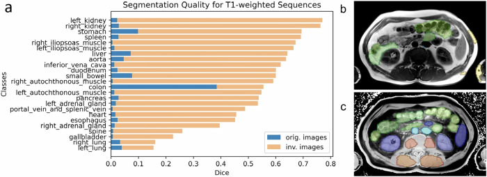
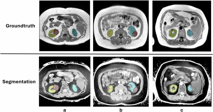
Results: Segmentation quality varied by MRI sequence and anatomical structure. Both models accurately segmented kidneys in T2wfs sequences without preprocessing (TotalSegmentator DSC 0.60), but TotalSegmentator failed to segment blood vessels and muscles. In T1w sequences, intensity inversion significantly improved TotalSegmentator performance, increasing the mean DSC across 24 structures from 0.04 to 0.56 (p < 0.001). Kidney tumor segmentation demonstrated poor performance in T2wfs sequences regardless of preprocessing. In T1w sequences, inversion improved tumor segmentation DSC from 0.04 to 0.42 (p < 0.001).
Conclusion: CT-trained models can generalize to MRI when supported by image augmentation. Inversion preprocessing enabled segmentation of renal cell carcinoma in T1w MRI using a CT-trained model. CT models might be transferable to the MRI domain.
Relevance statement: CT-trained artificial intelligence models can be adapted for MRI segmentation using simple preprocessing, potentially reducing manual annotation efforts and accelerating the development of AI-assisted tools for MRI analysis in research and future clinical practice.
Key points: CT segmentation models can create presegmentations for many structures in MRI scans. T1w MRI scans require an additional inversion step before segmenting with a CT model. Results were consistent for a large multiclass model (i.e., TotalSegmentator) and a smaller model for renal cell carcinoma.

 求助内容:
求助内容: 应助结果提醒方式:


