Kapil Kumar, Thomas Hartig Braunstein, Pablo Hernandez-Varas, Mikkel Baldtzer Liisberg and Bo W. Laursen
{"title":"一种水溶性、长荧光寿命DNA探针,用于活细胞有丝分裂的实时动态可视化,并适用于FLIM/时间门控成像。","authors":"Kapil Kumar, Thomas Hartig Braunstein, Pablo Hernandez-Varas, Mikkel Baldtzer Liisberg and Bo W. Laursen","doi":"10.1039/D5TB00404G","DOIUrl":null,"url":null,"abstract":"<p >Long-lived and photostable fluorescent probes for real-time monitoring of cell division, particularly for dynamic visualization of mitosis and interphase, are rare. We have developed a water-soluble diazaoxatriangulenium cation-based fluorescent probe, <strong>Nuc-DAOTA<small><sup>+</sup></small></strong>, that meets these criteria. <strong>Nuc-DAOTA<small><sup>+</sup></small></strong> has a long fluorescence lifetime (∼20 ns) and exclusively targets the nucleus for specific DNA binding in live cells, essential for real-time dynamic imaging of cell-division, including mitosis and interphase. Addition of dsDNA (0–50 μg mL<small><sup>−1</sup></small>) to <strong>Nuc-DAOTA<small><sup>+</sup></small></strong> results in a red shift (∼10 nm) of the absorption and a blue shift (∼5 nm) of the fluorescence maxima, along with an intensity increase in both. A very good linear correlation (<em>R</em><small><sup>2</sup></small> = 0.999) from the plot of fluorescence intensity <em>versus</em> concentration of dsDNA (up to 30 μg mL<small><sup>−1</sup></small>) resulted in a detection limit of 0.7 μg mL<small><sup>−1</sup></small>. The Benesi–Hildebrand plot was used to calculate the binding constant between the <strong>Nuc-DAOTA<small><sup>+</sup></small></strong> and DNA, which was found to be 1.6 × 10<small><sup>4</sup></small> M<small><sup>−1</sup></small> and the mechanism of binding interactions was investigated using CD spectroscopy. The long fluorescence lifetime and excellent biocompatibility of <strong>Nuc-DAOTA<small><sup>+</sup></small></strong> enabled its use for real-time dynamic imaging of mitotic phases and interphase in live CHO (Chinese hamster ovary) cells by using confocal microscopy. In addition, the <strong>Nuc-DAOTA<small><sup>+</sup></small></strong> exhibited high photostability during photo-bleaching experiments and was successfully applied for fluorescence lifetime imaging microscopy (FLIM) and time gated imaging of the mouse embryonic fibroblast 3T3 cell line.</p>","PeriodicalId":83,"journal":{"name":"Journal of Materials Chemistry B","volume":" 40","pages":" 13062-13074"},"PeriodicalIF":6.1000,"publicationDate":"2025-08-28","publicationTypes":"Journal Article","fieldsOfStudy":null,"isOpenAccess":false,"openAccessPdf":"","citationCount":"0","resultStr":"{\"title\":\"A water-soluble, long fluorescent lifetime DNA probe for real-time dynamic visualization of mitosis in live cells and applicability for FLIM/time-gated imaging\",\"authors\":\"Kapil Kumar, Thomas Hartig Braunstein, Pablo Hernandez-Varas, Mikkel Baldtzer Liisberg and Bo W. Laursen\",\"doi\":\"10.1039/D5TB00404G\",\"DOIUrl\":null,\"url\":null,\"abstract\":\"<p >Long-lived and photostable fluorescent probes for real-time monitoring of cell division, particularly for dynamic visualization of mitosis and interphase, are rare. We have developed a water-soluble diazaoxatriangulenium cation-based fluorescent probe, <strong>Nuc-DAOTA<small><sup>+</sup></small></strong>, that meets these criteria. <strong>Nuc-DAOTA<small><sup>+</sup></small></strong> has a long fluorescence lifetime (∼20 ns) and exclusively targets the nucleus for specific DNA binding in live cells, essential for real-time dynamic imaging of cell-division, including mitosis and interphase. Addition of dsDNA (0–50 μg mL<small><sup>−1</sup></small>) to <strong>Nuc-DAOTA<small><sup>+</sup></small></strong> results in a red shift (∼10 nm) of the absorption and a blue shift (∼5 nm) of the fluorescence maxima, along with an intensity increase in both. A very good linear correlation (<em>R</em><small><sup>2</sup></small> = 0.999) from the plot of fluorescence intensity <em>versus</em> concentration of dsDNA (up to 30 μg mL<small><sup>−1</sup></small>) resulted in a detection limit of 0.7 μg mL<small><sup>−1</sup></small>. The Benesi–Hildebrand plot was used to calculate the binding constant between the <strong>Nuc-DAOTA<small><sup>+</sup></small></strong> and DNA, which was found to be 1.6 × 10<small><sup>4</sup></small> M<small><sup>−1</sup></small> and the mechanism of binding interactions was investigated using CD spectroscopy. The long fluorescence lifetime and excellent biocompatibility of <strong>Nuc-DAOTA<small><sup>+</sup></small></strong> enabled its use for real-time dynamic imaging of mitotic phases and interphase in live CHO (Chinese hamster ovary) cells by using confocal microscopy. In addition, the <strong>Nuc-DAOTA<small><sup>+</sup></small></strong> exhibited high photostability during photo-bleaching experiments and was successfully applied for fluorescence lifetime imaging microscopy (FLIM) and time gated imaging of the mouse embryonic fibroblast 3T3 cell line.</p>\",\"PeriodicalId\":83,\"journal\":{\"name\":\"Journal of Materials Chemistry B\",\"volume\":\" 40\",\"pages\":\" 13062-13074\"},\"PeriodicalIF\":6.1000,\"publicationDate\":\"2025-08-28\",\"publicationTypes\":\"Journal Article\",\"fieldsOfStudy\":null,\"isOpenAccess\":false,\"openAccessPdf\":\"\",\"citationCount\":\"0\",\"resultStr\":null,\"platform\":\"Semanticscholar\",\"paperid\":null,\"PeriodicalName\":\"Journal of Materials Chemistry B\",\"FirstCategoryId\":\"1\",\"ListUrlMain\":\"https://pubs.rsc.org/en/content/articlelanding/2025/tb/d5tb00404g\",\"RegionNum\":3,\"RegionCategory\":\"医学\",\"ArticlePicture\":[],\"TitleCN\":null,\"AbstractTextCN\":null,\"PMCID\":null,\"EPubDate\":\"\",\"PubModel\":\"\",\"JCR\":\"Q1\",\"JCRName\":\"MATERIALS SCIENCE, BIOMATERIALS\",\"Score\":null,\"Total\":0}","platform":"Semanticscholar","paperid":null,"PeriodicalName":"Journal of Materials Chemistry B","FirstCategoryId":"1","ListUrlMain":"https://pubs.rsc.org/en/content/articlelanding/2025/tb/d5tb00404g","RegionNum":3,"RegionCategory":"医学","ArticlePicture":[],"TitleCN":null,"AbstractTextCN":null,"PMCID":null,"EPubDate":"","PubModel":"","JCR":"Q1","JCRName":"MATERIALS SCIENCE, BIOMATERIALS","Score":null,"Total":0}
A water-soluble, long fluorescent lifetime DNA probe for real-time dynamic visualization of mitosis in live cells and applicability for FLIM/time-gated imaging
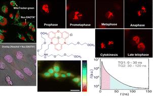
Long-lived and photostable fluorescent probes for real-time monitoring of cell division, particularly for dynamic visualization of mitosis and interphase, are rare. We have developed a water-soluble diazaoxatriangulenium cation-based fluorescent probe, Nuc-DAOTA+, that meets these criteria. Nuc-DAOTA+ has a long fluorescence lifetime (∼20 ns) and exclusively targets the nucleus for specific DNA binding in live cells, essential for real-time dynamic imaging of cell-division, including mitosis and interphase. Addition of dsDNA (0–50 μg mL−1) to Nuc-DAOTA+ results in a red shift (∼10 nm) of the absorption and a blue shift (∼5 nm) of the fluorescence maxima, along with an intensity increase in both. A very good linear correlation (R2 = 0.999) from the plot of fluorescence intensity versus concentration of dsDNA (up to 30 μg mL−1) resulted in a detection limit of 0.7 μg mL−1. The Benesi–Hildebrand plot was used to calculate the binding constant between the Nuc-DAOTA+ and DNA, which was found to be 1.6 × 10⁴ M−1 and the mechanism of binding interactions was investigated using CD spectroscopy. The long fluorescence lifetime and excellent biocompatibility of Nuc-DAOTA+ enabled its use for real-time dynamic imaging of mitotic phases and interphase in live CHO (Chinese hamster ovary) cells by using confocal microscopy. In addition, the Nuc-DAOTA+ exhibited high photostability during photo-bleaching experiments and was successfully applied for fluorescence lifetime imaging microscopy (FLIM) and time gated imaging of the mouse embryonic fibroblast 3T3 cell line.
期刊介绍:
Journal of Materials Chemistry A, B & C cover high quality studies across all fields of materials chemistry. The journals focus on those theoretical or experimental studies that report new understanding, applications, properties and synthesis of materials. Journal of Materials Chemistry A, B & C are separated by the intended application of the material studied. Broadly, applications in energy and sustainability are of interest to Journal of Materials Chemistry A, applications in biology and medicine are of interest to Journal of Materials Chemistry B, and applications in optical, magnetic and electronic devices are of interest to Journal of Materials Chemistry C.Journal of Materials Chemistry B is a Transformative Journal and Plan S compliant. Example topic areas within the scope of Journal of Materials Chemistry B are listed below. This list is neither exhaustive nor exclusive:
Antifouling coatings
Biocompatible materials
Bioelectronics
Bioimaging
Biomimetics
Biomineralisation
Bionics
Biosensors
Diagnostics
Drug delivery
Gene delivery
Immunobiology
Nanomedicine
Regenerative medicine & Tissue engineering
Scaffolds
Soft robotics
Stem cells
Therapeutic devices
 求助内容:
求助内容: 应助结果提醒方式:


