Hyeonji Sim, Yoojin An, Sung-Soo Kim, Danbee Kwon, Jeongmin Lee, Kichang Lee, Hakyoung Yoon
{"title":"猫健康与病变肾脏的肾皮质厚度与主动脉直径之比:超声对比评价。","authors":"Hyeonji Sim, Yoojin An, Sung-Soo Kim, Danbee Kwon, Jeongmin Lee, Kichang Lee, Hakyoung Yoon","doi":"10.1111/vru.70090","DOIUrl":null,"url":null,"abstract":"<p><p>In this retrospective multicenter study, we aimed to establish the renal cortical thickness-aortic diameter (RCT:Ao) ratio as a diagnostic parameter for detecting feline acute kidney injury or chronic kidney disease (AKI or CKD). This study included bilateral kidneys of 152 normal, 171 CKD, 19 AKI, and 15 acute-on-chronic kidney disease (ACKD) cats. Ultrasonographic measurements were obtained in the midsagittal plane of the kidneys and aorta. Multiple linear regression analysis of RCT, body weight (BW), and body condition score (BCS) revealed a positive correlation of RCT with BW (p < 0.001), but not with BCS (p = 0.343). Multiple linear regression analysis of RCT:Ao ratio, BW, and BCS showed a poor model fit (F value: 0.119). There were significant intergroup differences among the normal, CKD, AKI, and ACKD sub-cohorts (p < 0.001). Compared to normal cats, CKD and AKI cats each had lower and higher RCT:Ao ratio (both p < 0.001), respectively. The RCT:Ao ratio of the ACKD group significantly differed from that in normal and CKD groups (both p < 0.001), but not the AKI group (p = 0.159). Optimal RCT:Ao ratio cutoffs of 1.15 and 1.45 were used to distinguish between the normal and CKD groups (75% sensitivity, 80% specificity) and the normal and AKI groups (90% sensitivity, 89% specificity), respectively. The RCT:Ao ratio was unaffected by the BW and BCS and is a clinically useful diagnostic parameter for feline kidney disease.</p>","PeriodicalId":23581,"journal":{"name":"Veterinary Radiology & Ultrasound","volume":"66 5","pages":"e70090"},"PeriodicalIF":1.5000,"publicationDate":"2025-09-01","publicationTypes":"Journal Article","fieldsOfStudy":null,"isOpenAccess":false,"openAccessPdf":"https://www.ncbi.nlm.nih.gov/pmc/articles/PMC12445254/pdf/","citationCount":"0","resultStr":"{\"title\":\"Feline Renal Cortical Thickness-Aortic Diameter Ratio in Healthy Versus Diseased Kidneys: Comparative Ultrasonographic Evaluation.\",\"authors\":\"Hyeonji Sim, Yoojin An, Sung-Soo Kim, Danbee Kwon, Jeongmin Lee, Kichang Lee, Hakyoung Yoon\",\"doi\":\"10.1111/vru.70090\",\"DOIUrl\":null,\"url\":null,\"abstract\":\"<p><p>In this retrospective multicenter study, we aimed to establish the renal cortical thickness-aortic diameter (RCT:Ao) ratio as a diagnostic parameter for detecting feline acute kidney injury or chronic kidney disease (AKI or CKD). This study included bilateral kidneys of 152 normal, 171 CKD, 19 AKI, and 15 acute-on-chronic kidney disease (ACKD) cats. Ultrasonographic measurements were obtained in the midsagittal plane of the kidneys and aorta. Multiple linear regression analysis of RCT, body weight (BW), and body condition score (BCS) revealed a positive correlation of RCT with BW (p < 0.001), but not with BCS (p = 0.343). Multiple linear regression analysis of RCT:Ao ratio, BW, and BCS showed a poor model fit (F value: 0.119). There were significant intergroup differences among the normal, CKD, AKI, and ACKD sub-cohorts (p < 0.001). Compared to normal cats, CKD and AKI cats each had lower and higher RCT:Ao ratio (both p < 0.001), respectively. The RCT:Ao ratio of the ACKD group significantly differed from that in normal and CKD groups (both p < 0.001), but not the AKI group (p = 0.159). Optimal RCT:Ao ratio cutoffs of 1.15 and 1.45 were used to distinguish between the normal and CKD groups (75% sensitivity, 80% specificity) and the normal and AKI groups (90% sensitivity, 89% specificity), respectively. The RCT:Ao ratio was unaffected by the BW and BCS and is a clinically useful diagnostic parameter for feline kidney disease.</p>\",\"PeriodicalId\":23581,\"journal\":{\"name\":\"Veterinary Radiology & Ultrasound\",\"volume\":\"66 5\",\"pages\":\"e70090\"},\"PeriodicalIF\":1.5000,\"publicationDate\":\"2025-09-01\",\"publicationTypes\":\"Journal Article\",\"fieldsOfStudy\":null,\"isOpenAccess\":false,\"openAccessPdf\":\"https://www.ncbi.nlm.nih.gov/pmc/articles/PMC12445254/pdf/\",\"citationCount\":\"0\",\"resultStr\":null,\"platform\":\"Semanticscholar\",\"paperid\":null,\"PeriodicalName\":\"Veterinary Radiology & Ultrasound\",\"FirstCategoryId\":\"97\",\"ListUrlMain\":\"https://doi.org/10.1111/vru.70090\",\"RegionNum\":2,\"RegionCategory\":\"农林科学\",\"ArticlePicture\":[],\"TitleCN\":null,\"AbstractTextCN\":null,\"PMCID\":null,\"EPubDate\":\"\",\"PubModel\":\"\",\"JCR\":\"Q2\",\"JCRName\":\"VETERINARY SCIENCES\",\"Score\":null,\"Total\":0}","platform":"Semanticscholar","paperid":null,"PeriodicalName":"Veterinary Radiology & Ultrasound","FirstCategoryId":"97","ListUrlMain":"https://doi.org/10.1111/vru.70090","RegionNum":2,"RegionCategory":"农林科学","ArticlePicture":[],"TitleCN":null,"AbstractTextCN":null,"PMCID":null,"EPubDate":"","PubModel":"","JCR":"Q2","JCRName":"VETERINARY SCIENCES","Score":null,"Total":0}
Feline Renal Cortical Thickness-Aortic Diameter Ratio in Healthy Versus Diseased Kidneys: Comparative Ultrasonographic Evaluation.
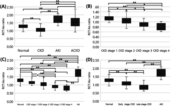
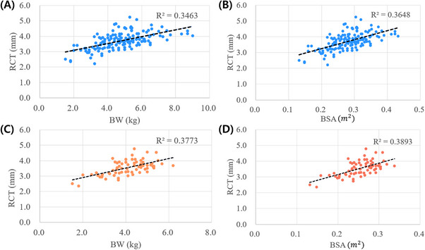
In this retrospective multicenter study, we aimed to establish the renal cortical thickness-aortic diameter (RCT:Ao) ratio as a diagnostic parameter for detecting feline acute kidney injury or chronic kidney disease (AKI or CKD). This study included bilateral kidneys of 152 normal, 171 CKD, 19 AKI, and 15 acute-on-chronic kidney disease (ACKD) cats. Ultrasonographic measurements were obtained in the midsagittal plane of the kidneys and aorta. Multiple linear regression analysis of RCT, body weight (BW), and body condition score (BCS) revealed a positive correlation of RCT with BW (p < 0.001), but not with BCS (p = 0.343). Multiple linear regression analysis of RCT:Ao ratio, BW, and BCS showed a poor model fit (F value: 0.119). There were significant intergroup differences among the normal, CKD, AKI, and ACKD sub-cohorts (p < 0.001). Compared to normal cats, CKD and AKI cats each had lower and higher RCT:Ao ratio (both p < 0.001), respectively. The RCT:Ao ratio of the ACKD group significantly differed from that in normal and CKD groups (both p < 0.001), but not the AKI group (p = 0.159). Optimal RCT:Ao ratio cutoffs of 1.15 and 1.45 were used to distinguish between the normal and CKD groups (75% sensitivity, 80% specificity) and the normal and AKI groups (90% sensitivity, 89% specificity), respectively. The RCT:Ao ratio was unaffected by the BW and BCS and is a clinically useful diagnostic parameter for feline kidney disease.
期刊介绍:
Veterinary Radiology & Ultrasound is a bimonthly, international, peer-reviewed, research journal devoted to the fields of veterinary diagnostic imaging and radiation oncology. Established in 1958, it is owned by the American College of Veterinary Radiology and is also the official journal for six affiliate veterinary organizations. Veterinary Radiology & Ultrasound is represented on the International Committee of Medical Journal Editors, World Association of Medical Editors, and Committee on Publication Ethics.
The mission of Veterinary Radiology & Ultrasound is to serve as a leading resource for high quality articles that advance scientific knowledge and standards of clinical practice in the areas of veterinary diagnostic radiology, computed tomography, magnetic resonance imaging, ultrasonography, nuclear imaging, radiation oncology, and interventional radiology. Manuscript types include original investigations, imaging diagnosis reports, review articles, editorials and letters to the Editor. Acceptance criteria include originality, significance, quality, reader interest, composition and adherence to author guidelines.

 求助内容:
求助内容: 应助结果提醒方式:


