一新种环棘虫南通n. Gen. n. Sp.的形态和分子特征。
IF 2.4
3区 生物学
Q1 ZOOLOGY
引用次数: 0
摘要
摘要从江苏南通市富营养化水体中分离到一种新的小孢子虫Cyclopispore南通n. gen. n. sp.。由于大量孢子积聚在脂肪组织中,感染的桡足类动物通常表现为不透明。最早观察到的发育阶段是与宿主细胞质直接接触的无核小体。成熟孢子为梨形单核,长4.38 ± 0.20(4.14-4.81)µm,宽2.94 ± 0.09(2.78-3.12)µm。双侧极质体由宽的前片和紧密排列的后片组成。等丝极性灯丝卷成13-16圈,排列成3-4排。得到的1306 bp的SSU序列与所鉴定的细孢子虫AY09006的相似性最高(85.9 %)。系统发育分析表明,本种与一个未确定的具有高支持值的感染黑桫椤的种聚集在一起,形成了一个独立的分支,定位于glomerata- gurleya - agglomerata - binucleata - senoma - pseudoberwaldia谱系的基础。根据其形态特征和超微结构特征,以及SSU rdna推断的系统发育关系,我们提出了一个新的属(Cyclopispore n. gen.)和种(Cyclopispore n通n. sp.)来包含该寄生虫。本文章由计算机程序翻译,如有差异,请以英文原文为准。
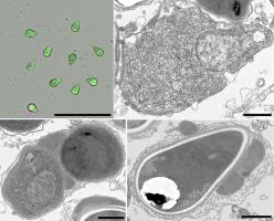
Morphological and molecular characterization of a new species, Cyclopispora nantong n. gen. n. sp. from the adipose tissue of Eucyclops speratus (Crustacea: Cyclopidae)
A new microsporidium, Cyclopispora nantong n. gen. n. sp., was described from the adipose tissue of Eucyclops speratus collected from a eutrophic water body of Nantong city, Jiangsu province, China. Infected copepods generally appeared opaque due to numerous spores accumulated in the adipose tissue. The earliest developmental stages observed were uninucleate meronts which resided in direct contact with the host cell cytoplasm. Mature spores were pyriform, monokaryotic, and measured 4.38 ± 0.20 (4.14–4.81) µm in length and 2.94 ± 0.09 (2.78–3.12) µm in width. The bipartite polaroplast consisted of the wide anterior lamellae and the tightly packed posterior lamellae. Isofilar polar filament coiled 13–16 turns and arranged in 3–4 rows. The 1306 bp SSU sequence obtained from E. speratus showed the highest similarity (85.9 %) to Hazardia milleri (AY09006) among the identified microsporidia. Phylogenetic analysis indicated that the present species clustered with an unidentified B. latissima lata-infecting species with high support values to form an independent branch which positioned the base of the Conglomerata-Gurleya-Agglomerata-Binucleata-Senoma-Pseudoberwaldia lineage. Based on the morphological characters and ultrastructural features, as well as SSU rDNA-inferred phylogenetic relationships, we propose the establishment of a novel genus (Cyclopispora n. gen.) and species (Cyclopispora nantong n. sp.) to contain this parasite.
求助全文
通过发布文献求助,成功后即可免费获取论文全文。
去求助
来源期刊
CiteScore
6.10
自引率
5.90%
发文量
94
审稿时长
1 months
期刊介绍:
The Journal of Invertebrate Pathology presents original research articles and notes on the induction and pathogenesis of diseases of invertebrates, including the suppression of diseases in beneficial species, and the use of diseases in controlling undesirable species. In addition, the journal publishes the results of physiological, morphological, genetic, immunological and ecological studies as related to the etiologic agents of diseases of invertebrates.
The Journal of Invertebrate Pathology is the adopted journal of the Society for Invertebrate Pathology, and is available to SIP members at a special reduced price.
 求助内容:
求助内容: 应助结果提醒方式:


