Andrei Bancu, Matthew Hanks, Lauren Ackroyd, Suresh Venkatachalapathy, Dileep N Lobo, Abed M Zaitoun
{"title":"胰腺囊性病变的细胞学和核心活检:三种细胞学分级系统的比较。","authors":"Andrei Bancu, Matthew Hanks, Lauren Ackroyd, Suresh Venkatachalapathy, Dileep N Lobo, Abed M Zaitoun","doi":"10.4103/joc.joc_6_25","DOIUrl":null,"url":null,"abstract":"<p><strong>Introduction: </strong>Cytological appearances may be insufficient to establish the diagnosis of pancreatic cystic lesions, especially for premalignant neoplasms. This study aimed to evaluate three commonly used cytological grading systems [C1-C5, Papanicolaou (Pap), and World Health Organization (WHO) classification systems] in assessing pancreatic cystic lesions.</p><p><strong>Materials and methods: </strong>A total of 210 pancreatic cytology specimens were classified based on the aforementioned grading systems, 127 of which had supporting histology reported across 6 years, at a single-tertiary referral center.</p><p><strong>Results: </strong>We excluded 26 cases because of inadequate cytology. The most common cystic lesions were intraductal papillary mucinous neoplasms (IPMNs, <i>n</i> = 71) and pseudocysts (<i>n</i> = 55). Among IPMN, 27 were moderate/high-grade, and 44 were low-grade. There were 12 mucinous cystic neoplasms, 75 benign cysts (pseudocysts, serous cystadenomas, lymphoepithelial cysts, and others), and 15 malignant cases, with 11 cysts being of uncertain etiology. There were 21 high-risk and potentially malignant (WHO grade V) cases in comparison with 42 cases using the C grading (C4 and C5). However, according to Pap grading, 87 had varying risks of malignancy (Category IVB). According to the WHO classification, 63 cases were classified as low-risk pancreaticobiliary neoplasms (WHO grade IV). The remaining cases 85 were benign to very low-risk malignant potential and, therefore, were less likely to be considered for surgical intervention. Diagnostic concordance was higher between cytology and core biopsies in low-grade IPMN.</p><p><strong>Conclusion: </strong>The WHO system provides better risk stratification for neoplasms, optimizing surgical management. However, the C1-C5 system does not recognize cystic lesions with malignant potential.</p>","PeriodicalId":50217,"journal":{"name":"Journal of Cytology","volume":"42 3","pages":"151-162"},"PeriodicalIF":1.0000,"publicationDate":"2025-07-01","publicationTypes":"Journal Article","fieldsOfStudy":null,"isOpenAccess":false,"openAccessPdf":"https://www.ncbi.nlm.nih.gov/pmc/articles/PMC12435879/pdf/","citationCount":"0","resultStr":"{\"title\":\"Cytology and Core Biopsy for Cystic Lesions of the Pancreas: A Comparison of Three Cytological Grading Systems.\",\"authors\":\"Andrei Bancu, Matthew Hanks, Lauren Ackroyd, Suresh Venkatachalapathy, Dileep N Lobo, Abed M Zaitoun\",\"doi\":\"10.4103/joc.joc_6_25\",\"DOIUrl\":null,\"url\":null,\"abstract\":\"<p><strong>Introduction: </strong>Cytological appearances may be insufficient to establish the diagnosis of pancreatic cystic lesions, especially for premalignant neoplasms. This study aimed to evaluate three commonly used cytological grading systems [C1-C5, Papanicolaou (Pap), and World Health Organization (WHO) classification systems] in assessing pancreatic cystic lesions.</p><p><strong>Materials and methods: </strong>A total of 210 pancreatic cytology specimens were classified based on the aforementioned grading systems, 127 of which had supporting histology reported across 6 years, at a single-tertiary referral center.</p><p><strong>Results: </strong>We excluded 26 cases because of inadequate cytology. The most common cystic lesions were intraductal papillary mucinous neoplasms (IPMNs, <i>n</i> = 71) and pseudocysts (<i>n</i> = 55). Among IPMN, 27 were moderate/high-grade, and 44 were low-grade. There were 12 mucinous cystic neoplasms, 75 benign cysts (pseudocysts, serous cystadenomas, lymphoepithelial cysts, and others), and 15 malignant cases, with 11 cysts being of uncertain etiology. There were 21 high-risk and potentially malignant (WHO grade V) cases in comparison with 42 cases using the C grading (C4 and C5). However, according to Pap grading, 87 had varying risks of malignancy (Category IVB). According to the WHO classification, 63 cases were classified as low-risk pancreaticobiliary neoplasms (WHO grade IV). The remaining cases 85 were benign to very low-risk malignant potential and, therefore, were less likely to be considered for surgical intervention. Diagnostic concordance was higher between cytology and core biopsies in low-grade IPMN.</p><p><strong>Conclusion: </strong>The WHO system provides better risk stratification for neoplasms, optimizing surgical management. However, the C1-C5 system does not recognize cystic lesions with malignant potential.</p>\",\"PeriodicalId\":50217,\"journal\":{\"name\":\"Journal of Cytology\",\"volume\":\"42 3\",\"pages\":\"151-162\"},\"PeriodicalIF\":1.0000,\"publicationDate\":\"2025-07-01\",\"publicationTypes\":\"Journal Article\",\"fieldsOfStudy\":null,\"isOpenAccess\":false,\"openAccessPdf\":\"https://www.ncbi.nlm.nih.gov/pmc/articles/PMC12435879/pdf/\",\"citationCount\":\"0\",\"resultStr\":null,\"platform\":\"Semanticscholar\",\"paperid\":null,\"PeriodicalName\":\"Journal of Cytology\",\"FirstCategoryId\":\"3\",\"ListUrlMain\":\"https://doi.org/10.4103/joc.joc_6_25\",\"RegionNum\":4,\"RegionCategory\":\"医学\",\"ArticlePicture\":[],\"TitleCN\":null,\"AbstractTextCN\":null,\"PMCID\":null,\"EPubDate\":\"2025/8/29 0:00:00\",\"PubModel\":\"Epub\",\"JCR\":\"Q4\",\"JCRName\":\"MEDICAL LABORATORY TECHNOLOGY\",\"Score\":null,\"Total\":0}","platform":"Semanticscholar","paperid":null,"PeriodicalName":"Journal of Cytology","FirstCategoryId":"3","ListUrlMain":"https://doi.org/10.4103/joc.joc_6_25","RegionNum":4,"RegionCategory":"医学","ArticlePicture":[],"TitleCN":null,"AbstractTextCN":null,"PMCID":null,"EPubDate":"2025/8/29 0:00:00","PubModel":"Epub","JCR":"Q4","JCRName":"MEDICAL LABORATORY TECHNOLOGY","Score":null,"Total":0}
Cytology and Core Biopsy for Cystic Lesions of the Pancreas: A Comparison of Three Cytological Grading Systems.
Introduction: Cytological appearances may be insufficient to establish the diagnosis of pancreatic cystic lesions, especially for premalignant neoplasms. This study aimed to evaluate three commonly used cytological grading systems [C1-C5, Papanicolaou (Pap), and World Health Organization (WHO) classification systems] in assessing pancreatic cystic lesions.
Materials and methods: A total of 210 pancreatic cytology specimens were classified based on the aforementioned grading systems, 127 of which had supporting histology reported across 6 years, at a single-tertiary referral center.
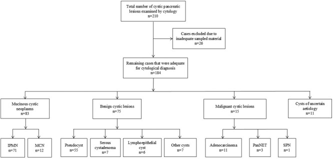
Results: We excluded 26 cases because of inadequate cytology. The most common cystic lesions were intraductal papillary mucinous neoplasms (IPMNs, n = 71) and pseudocysts (n = 55). Among IPMN, 27 were moderate/high-grade, and 44 were low-grade. There were 12 mucinous cystic neoplasms, 75 benign cysts (pseudocysts, serous cystadenomas, lymphoepithelial cysts, and others), and 15 malignant cases, with 11 cysts being of uncertain etiology. There were 21 high-risk and potentially malignant (WHO grade V) cases in comparison with 42 cases using the C grading (C4 and C5). However, according to Pap grading, 87 had varying risks of malignancy (Category IVB). According to the WHO classification, 63 cases were classified as low-risk pancreaticobiliary neoplasms (WHO grade IV). The remaining cases 85 were benign to very low-risk malignant potential and, therefore, were less likely to be considered for surgical intervention. Diagnostic concordance was higher between cytology and core biopsies in low-grade IPMN.
Conclusion: The WHO system provides better risk stratification for neoplasms, optimizing surgical management. However, the C1-C5 system does not recognize cystic lesions with malignant potential.
期刊介绍:
The Journal of Cytology is the official Quarterly publication of the Indian Academy of Cytologists. It is in the 25th year of publication in the year 2008. The journal covers all aspects of diagnostic cytology, including fine needle aspiration cytology, gynecological and non-gynecological cytology. Articles on ancillary techniques, like cytochemistry, immunocytochemistry, electron microscopy, molecular cytopathology, as applied to cytological material are also welcome. The journal gives preference to clinically oriented studies over experimental and animal studies. The Journal would publish peer-reviewed original research papers, case reports, systematic reviews, meta-analysis, and debates.
 求助内容:
求助内容: 应助结果提醒方式:


