{"title":"尿细胞学的机器学习强调肌肉侵袭性尿路上皮癌中中性粒细胞计数增加。","authors":"Moe Kameda, Sayaka Kobayashi, Yoshimi Nishijima, Ryosuke Akuzawa, Rio Kaneko, Rio Shibanuma, Seiji Arai, Hayato Ikota, Kazuhiro Suzuki, Hideaki Yokoo, Masanao Saio","doi":"10.4103/joc.joc_158_24","DOIUrl":null,"url":null,"abstract":"<p><strong>Objective: </strong>This study conducted an unsupervised learning cluster analysis on urine cytological images of high-grade urothelial carcinoma to assess their explanatory potential.</p><p><strong>Materials and methods: </strong>A total of 124 urine cytology specimens of urothelial carcinoma, collected between December 2010 to December 2021 at Gunma University Hospital, were analyzed. Ten cytological image fields per specimen were captured, and pathological T factors were examined using principal component analysis and t-distributed stochastic neighbor embedding (t-SNE) with machine learning (ML) software. Common image features were also verbalized and manually reevaluated.</p><p><strong>Results: </strong>In the t-SNE analysis, the T1-dominant region was characterized by \"few cells in the background,\" whereas the T2-dominant region showed \"many cells in the image,\" \"numerous neutrophils in the image,\" and \"abundant tumor cells in the image.\" Human reassessment identified significant differences related to muscle invasion status for all findings except \"abundant tumor cells in the image.\" Furthermore, we confirmed that histological neutrophil infiltration was related to the abundance of neutrophils in the cytological specimens.</p><p><strong>Conclusion: </strong>This study is noteworthy as the cluster analysis identified previously unreported variations in background cell types and quality linked to muscle invasion status, and it also demonstrated the explainability of ML-derived findings through manual reassessment.</p>","PeriodicalId":50217,"journal":{"name":"Journal of Cytology","volume":"42 3","pages":"124-133"},"PeriodicalIF":1.0000,"publicationDate":"2025-07-01","publicationTypes":"Journal Article","fieldsOfStudy":null,"isOpenAccess":false,"openAccessPdf":"https://www.ncbi.nlm.nih.gov/pmc/articles/PMC12435878/pdf/","citationCount":"0","resultStr":"{\"title\":\"Machine Learning of Urine Cytology Highlights Increased Neutrophil Count in Muscle-Invasive Urothelial Carcinoma.\",\"authors\":\"Moe Kameda, Sayaka Kobayashi, Yoshimi Nishijima, Ryosuke Akuzawa, Rio Kaneko, Rio Shibanuma, Seiji Arai, Hayato Ikota, Kazuhiro Suzuki, Hideaki Yokoo, Masanao Saio\",\"doi\":\"10.4103/joc.joc_158_24\",\"DOIUrl\":null,\"url\":null,\"abstract\":\"<p><strong>Objective: </strong>This study conducted an unsupervised learning cluster analysis on urine cytological images of high-grade urothelial carcinoma to assess their explanatory potential.</p><p><strong>Materials and methods: </strong>A total of 124 urine cytology specimens of urothelial carcinoma, collected between December 2010 to December 2021 at Gunma University Hospital, were analyzed. Ten cytological image fields per specimen were captured, and pathological T factors were examined using principal component analysis and t-distributed stochastic neighbor embedding (t-SNE) with machine learning (ML) software. Common image features were also verbalized and manually reevaluated.</p><p><strong>Results: </strong>In the t-SNE analysis, the T1-dominant region was characterized by \\\"few cells in the background,\\\" whereas the T2-dominant region showed \\\"many cells in the image,\\\" \\\"numerous neutrophils in the image,\\\" and \\\"abundant tumor cells in the image.\\\" Human reassessment identified significant differences related to muscle invasion status for all findings except \\\"abundant tumor cells in the image.\\\" Furthermore, we confirmed that histological neutrophil infiltration was related to the abundance of neutrophils in the cytological specimens.</p><p><strong>Conclusion: </strong>This study is noteworthy as the cluster analysis identified previously unreported variations in background cell types and quality linked to muscle invasion status, and it also demonstrated the explainability of ML-derived findings through manual reassessment.</p>\",\"PeriodicalId\":50217,\"journal\":{\"name\":\"Journal of Cytology\",\"volume\":\"42 3\",\"pages\":\"124-133\"},\"PeriodicalIF\":1.0000,\"publicationDate\":\"2025-07-01\",\"publicationTypes\":\"Journal Article\",\"fieldsOfStudy\":null,\"isOpenAccess\":false,\"openAccessPdf\":\"https://www.ncbi.nlm.nih.gov/pmc/articles/PMC12435878/pdf/\",\"citationCount\":\"0\",\"resultStr\":null,\"platform\":\"Semanticscholar\",\"paperid\":null,\"PeriodicalName\":\"Journal of Cytology\",\"FirstCategoryId\":\"3\",\"ListUrlMain\":\"https://doi.org/10.4103/joc.joc_158_24\",\"RegionNum\":4,\"RegionCategory\":\"医学\",\"ArticlePicture\":[],\"TitleCN\":null,\"AbstractTextCN\":null,\"PMCID\":null,\"EPubDate\":\"2025/8/29 0:00:00\",\"PubModel\":\"Epub\",\"JCR\":\"Q4\",\"JCRName\":\"MEDICAL LABORATORY TECHNOLOGY\",\"Score\":null,\"Total\":0}","platform":"Semanticscholar","paperid":null,"PeriodicalName":"Journal of Cytology","FirstCategoryId":"3","ListUrlMain":"https://doi.org/10.4103/joc.joc_158_24","RegionNum":4,"RegionCategory":"医学","ArticlePicture":[],"TitleCN":null,"AbstractTextCN":null,"PMCID":null,"EPubDate":"2025/8/29 0:00:00","PubModel":"Epub","JCR":"Q4","JCRName":"MEDICAL LABORATORY TECHNOLOGY","Score":null,"Total":0}
Machine Learning of Urine Cytology Highlights Increased Neutrophil Count in Muscle-Invasive Urothelial Carcinoma.
Objective: This study conducted an unsupervised learning cluster analysis on urine cytological images of high-grade urothelial carcinoma to assess their explanatory potential.
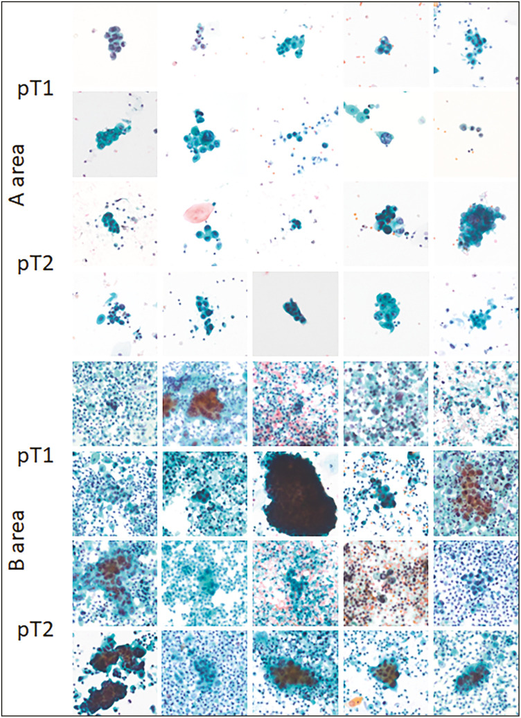
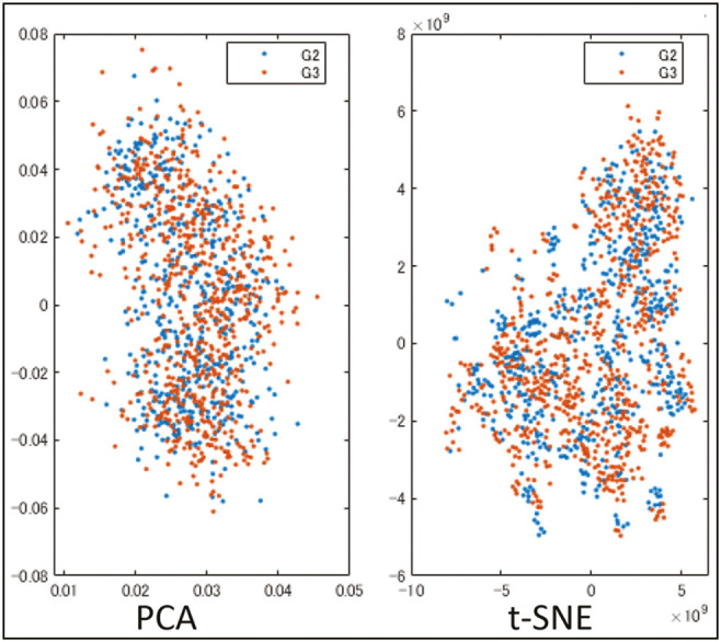
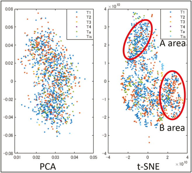
Materials and methods: A total of 124 urine cytology specimens of urothelial carcinoma, collected between December 2010 to December 2021 at Gunma University Hospital, were analyzed. Ten cytological image fields per specimen were captured, and pathological T factors were examined using principal component analysis and t-distributed stochastic neighbor embedding (t-SNE) with machine learning (ML) software. Common image features were also verbalized and manually reevaluated.
Results: In the t-SNE analysis, the T1-dominant region was characterized by "few cells in the background," whereas the T2-dominant region showed "many cells in the image," "numerous neutrophils in the image," and "abundant tumor cells in the image." Human reassessment identified significant differences related to muscle invasion status for all findings except "abundant tumor cells in the image." Furthermore, we confirmed that histological neutrophil infiltration was related to the abundance of neutrophils in the cytological specimens.
Conclusion: This study is noteworthy as the cluster analysis identified previously unreported variations in background cell types and quality linked to muscle invasion status, and it also demonstrated the explainability of ML-derived findings through manual reassessment.
期刊介绍:
The Journal of Cytology is the official Quarterly publication of the Indian Academy of Cytologists. It is in the 25th year of publication in the year 2008. The journal covers all aspects of diagnostic cytology, including fine needle aspiration cytology, gynecological and non-gynecological cytology. Articles on ancillary techniques, like cytochemistry, immunocytochemistry, electron microscopy, molecular cytopathology, as applied to cytological material are also welcome. The journal gives preference to clinically oriented studies over experimental and animal studies. The Journal would publish peer-reviewed original research papers, case reports, systematic reviews, meta-analysis, and debates.
 求助内容:
求助内容: 应助结果提醒方式:


