Almoayad M Makrami, Yazeed J Alhaqbani, Dhoha M Alhamad, Talaat J Hamdi, Abdulaziz S Khoshaim
{"title":"在蛇形脉络膜炎的标准治疗中加入玻璃体内地塞米松对快速缓解的作用:1例报告。","authors":"Almoayad M Makrami, Yazeed J Alhaqbani, Dhoha M Alhamad, Talaat J Hamdi, Abdulaziz S Khoshaim","doi":"10.2147/IMCRJ.S536828","DOIUrl":null,"url":null,"abstract":"<p><strong>Background: </strong>This case highlights the potential of combining intravitreal dexamethasone implants as first line of management with reduced dose of systemic steroid therapy to achieve rapid remission in acute serpiginous choroiditis. To prevent systemic adverse effects and obtain rapid control of serpiginous lesions, as necessary in the case presented, local therapy using a dexamethasone intravitreal implant may be considered as a complement to systemic treatment.</p><p><strong>Case presentation: </strong>A 42 year old woman with steadily declining vision in her right eye for the previous four months presented to the emergency department. On the Snellen chart, the best-corrected visual acuity was 20/28 in the left eye and 20/100 in the right eye. The intraocular pressure (IOP) of the right and left eyes was 15 and 16 mmHg, respectively. Anterior segment examination was unremarkable. Fundus examination of the right eye revealed a gray finger-like lesion with an active border. The left eye showed a small yellowish-finger-like lesion involving the nasal macula. Disruption in the ellipsoid zone in the right eye and the parafoveal region in the left eye was demonstrated using optical coherence tomography (OCT). Fundus fluorescein angiography revealed bilateral, finger-like branching lesions were seen on fundus fluorescein angiography (FFA). Fundus Autofluorescence (FAF) showed hypoautofluorescence lesions with hyper-autofluorescence edges. Both eyes were diagnosed with active serpiginous choroiditis, after excluding any active infections through blood work-up. The patient was started oral prednisolone 0.5 mg/kg tapering; however, because the patient would be traveling for three weeks, the systemic oral steroid with an intravitreal dexamethasone implant in each eye. After nine days, there was noticeable improvement in the visual acuity of the right eye with normal intraocular pressure. OCT showed minimal restoration of the ellipsoid zone in the right eye, with resolved inflammatory material in both eyes (Figure 1). After starting Azathioprine, disease activity was suppressed for six months without relapsing.</p><p><strong>Conclusion: </strong>This case raises a question about the benefit of combined therapy for quick vision restoration, inhibiting further destruction of outer retinal layers during the management of acute attack and reduction of overall systemic steroids dose together with its complications versus the risk of local steroids administration and cost-effectiveness. Additional research is required to validate this finding.</p>","PeriodicalId":14337,"journal":{"name":"International Medical Case Reports Journal","volume":"18 ","pages":"1205-1210"},"PeriodicalIF":0.7000,"publicationDate":"2025-09-11","publicationTypes":"Journal Article","fieldsOfStudy":null,"isOpenAccess":false,"openAccessPdf":"https://www.ncbi.nlm.nih.gov/pmc/articles/PMC12435371/pdf/","citationCount":"0","resultStr":"{\"title\":\"The Role of Adding Intravitreal Dexamethasone Implant to the Standard Management of Serpiginous Choroiditis for Achieving Rapid Remission: A Case Report.\",\"authors\":\"Almoayad M Makrami, Yazeed J Alhaqbani, Dhoha M Alhamad, Talaat J Hamdi, Abdulaziz S Khoshaim\",\"doi\":\"10.2147/IMCRJ.S536828\",\"DOIUrl\":null,\"url\":null,\"abstract\":\"<p><strong>Background: </strong>This case highlights the potential of combining intravitreal dexamethasone implants as first line of management with reduced dose of systemic steroid therapy to achieve rapid remission in acute serpiginous choroiditis. To prevent systemic adverse effects and obtain rapid control of serpiginous lesions, as necessary in the case presented, local therapy using a dexamethasone intravitreal implant may be considered as a complement to systemic treatment.</p><p><strong>Case presentation: </strong>A 42 year old woman with steadily declining vision in her right eye for the previous four months presented to the emergency department. On the Snellen chart, the best-corrected visual acuity was 20/28 in the left eye and 20/100 in the right eye. The intraocular pressure (IOP) of the right and left eyes was 15 and 16 mmHg, respectively. Anterior segment examination was unremarkable. Fundus examination of the right eye revealed a gray finger-like lesion with an active border. The left eye showed a small yellowish-finger-like lesion involving the nasal macula. Disruption in the ellipsoid zone in the right eye and the parafoveal region in the left eye was demonstrated using optical coherence tomography (OCT). Fundus fluorescein angiography revealed bilateral, finger-like branching lesions were seen on fundus fluorescein angiography (FFA). Fundus Autofluorescence (FAF) showed hypoautofluorescence lesions with hyper-autofluorescence edges. Both eyes were diagnosed with active serpiginous choroiditis, after excluding any active infections through blood work-up. The patient was started oral prednisolone 0.5 mg/kg tapering; however, because the patient would be traveling for three weeks, the systemic oral steroid with an intravitreal dexamethasone implant in each eye. After nine days, there was noticeable improvement in the visual acuity of the right eye with normal intraocular pressure. OCT showed minimal restoration of the ellipsoid zone in the right eye, with resolved inflammatory material in both eyes (Figure 1). After starting Azathioprine, disease activity was suppressed for six months without relapsing.</p><p><strong>Conclusion: </strong>This case raises a question about the benefit of combined therapy for quick vision restoration, inhibiting further destruction of outer retinal layers during the management of acute attack and reduction of overall systemic steroids dose together with its complications versus the risk of local steroids administration and cost-effectiveness. Additional research is required to validate this finding.</p>\",\"PeriodicalId\":14337,\"journal\":{\"name\":\"International Medical Case Reports Journal\",\"volume\":\"18 \",\"pages\":\"1205-1210\"},\"PeriodicalIF\":0.7000,\"publicationDate\":\"2025-09-11\",\"publicationTypes\":\"Journal Article\",\"fieldsOfStudy\":null,\"isOpenAccess\":false,\"openAccessPdf\":\"https://www.ncbi.nlm.nih.gov/pmc/articles/PMC12435371/pdf/\",\"citationCount\":\"0\",\"resultStr\":null,\"platform\":\"Semanticscholar\",\"paperid\":null,\"PeriodicalName\":\"International Medical Case Reports Journal\",\"FirstCategoryId\":\"1085\",\"ListUrlMain\":\"https://doi.org/10.2147/IMCRJ.S536828\",\"RegionNum\":0,\"RegionCategory\":null,\"ArticlePicture\":[],\"TitleCN\":null,\"AbstractTextCN\":null,\"PMCID\":null,\"EPubDate\":\"2025/1/1 0:00:00\",\"PubModel\":\"eCollection\",\"JCR\":\"Q3\",\"JCRName\":\"MEDICINE, GENERAL & INTERNAL\",\"Score\":null,\"Total\":0}","platform":"Semanticscholar","paperid":null,"PeriodicalName":"International Medical Case Reports Journal","FirstCategoryId":"1085","ListUrlMain":"https://doi.org/10.2147/IMCRJ.S536828","RegionNum":0,"RegionCategory":null,"ArticlePicture":[],"TitleCN":null,"AbstractTextCN":null,"PMCID":null,"EPubDate":"2025/1/1 0:00:00","PubModel":"eCollection","JCR":"Q3","JCRName":"MEDICINE, GENERAL & INTERNAL","Score":null,"Total":0}
The Role of Adding Intravitreal Dexamethasone Implant to the Standard Management of Serpiginous Choroiditis for Achieving Rapid Remission: A Case Report.
Background: This case highlights the potential of combining intravitreal dexamethasone implants as first line of management with reduced dose of systemic steroid therapy to achieve rapid remission in acute serpiginous choroiditis. To prevent systemic adverse effects and obtain rapid control of serpiginous lesions, as necessary in the case presented, local therapy using a dexamethasone intravitreal implant may be considered as a complement to systemic treatment.
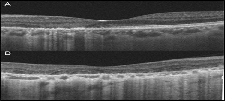
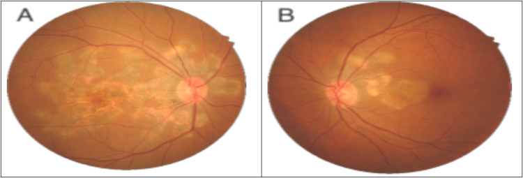
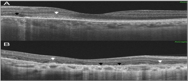
Case presentation: A 42 year old woman with steadily declining vision in her right eye for the previous four months presented to the emergency department. On the Snellen chart, the best-corrected visual acuity was 20/28 in the left eye and 20/100 in the right eye. The intraocular pressure (IOP) of the right and left eyes was 15 and 16 mmHg, respectively. Anterior segment examination was unremarkable. Fundus examination of the right eye revealed a gray finger-like lesion with an active border. The left eye showed a small yellowish-finger-like lesion involving the nasal macula. Disruption in the ellipsoid zone in the right eye and the parafoveal region in the left eye was demonstrated using optical coherence tomography (OCT). Fundus fluorescein angiography revealed bilateral, finger-like branching lesions were seen on fundus fluorescein angiography (FFA). Fundus Autofluorescence (FAF) showed hypoautofluorescence lesions with hyper-autofluorescence edges. Both eyes were diagnosed with active serpiginous choroiditis, after excluding any active infections through blood work-up. The patient was started oral prednisolone 0.5 mg/kg tapering; however, because the patient would be traveling for three weeks, the systemic oral steroid with an intravitreal dexamethasone implant in each eye. After nine days, there was noticeable improvement in the visual acuity of the right eye with normal intraocular pressure. OCT showed minimal restoration of the ellipsoid zone in the right eye, with resolved inflammatory material in both eyes (Figure 1). After starting Azathioprine, disease activity was suppressed for six months without relapsing.
Conclusion: This case raises a question about the benefit of combined therapy for quick vision restoration, inhibiting further destruction of outer retinal layers during the management of acute attack and reduction of overall systemic steroids dose together with its complications versus the risk of local steroids administration and cost-effectiveness. Additional research is required to validate this finding.
期刊介绍:
International Medical Case Reports Journal is an international, peer-reviewed, open access, online journal publishing original case reports from all medical specialties. Submissions should not normally exceed 3,000 words or 4 published pages including figures, diagrams and references. As of 1st April 2019, the International Medical Case Reports Journal will no longer consider meta-analyses for publication.

 求助内容:
求助内容: 应助结果提醒方式:


