Simmi Gupta, Esha S. Attar, Vishvesh Joshi and Padma V. Devarajan
{"title":"羟基磷灰石纳米颗粒形状对骨肉瘤细胞内化和抗癌效果的影响","authors":"Simmi Gupta, Esha S. Attar, Vishvesh Joshi and Padma V. Devarajan","doi":"10.1039/D5PM00005J","DOIUrl":null,"url":null,"abstract":"<p >Hydroxyapatite nanoparticles (HAP NPs) with distinct morphologies were synthesized by the wet precipitation method by varying pH, and their shape was confirmed by scanning electron microscopy as spherical (pH 12), rod-like (pH 9), and needle-like (pH 8). The particle sizes of HAP NPs were 96.86 ± 1.48 nm for needle-shaped, 118 ± 4.32 nm for rod-shaped, and 94.43 ± 1.02 nm for spherical-shaped particles. XRD analysis showed clear and distinct peaks indicating crystalline nature, while FTIR confirmed the characteristic features of hydroxyapatite. The negative zeta potential of the HAP NPs was attributed to the presence of surface phosphate ions. The influence of HAP NP shape and size on intracellular uptake was evaluated in the MG-63 osteosarcoma cell line by Confocal Laser Scanning Microscopy (CLSM). CLSM results demonstrated that rod-shaped HAP NPs predominantly localized within the lysosome and nucleus, while spherical HAP NPs accumulated at the cell membrane. The MTT, clonogenic survival, cell scratch and transwell migration assays revealed that rod-shaped HAP NPs exhibited superior anticancer activity compared to their needle- and spherical-shaped counterparts and completely suppressed the clonogenic survival of MG-63 cells. Our findings confirm that the shape of HAP NPs is a critical factor influencing their intracellular uptake and anticancer activity.</p>","PeriodicalId":101141,"journal":{"name":"RSC Pharmaceutics","volume":" 5","pages":" 1087-1095"},"PeriodicalIF":0.0000,"publicationDate":"2025-04-24","publicationTypes":"Journal Article","fieldsOfStudy":null,"isOpenAccess":false,"openAccessPdf":"https://pubs.rsc.org/en/content/articlepdf/2025/pm/d5pm00005j?page=search","citationCount":"0","resultStr":"{\"title\":\"Effect of shape on cellular internalization and anti-cancer efficacy of hydroxyapatite nanoparticles in an osteosarcoma cell line†\",\"authors\":\"Simmi Gupta, Esha S. Attar, Vishvesh Joshi and Padma V. Devarajan\",\"doi\":\"10.1039/D5PM00005J\",\"DOIUrl\":null,\"url\":null,\"abstract\":\"<p >Hydroxyapatite nanoparticles (HAP NPs) with distinct morphologies were synthesized by the wet precipitation method by varying pH, and their shape was confirmed by scanning electron microscopy as spherical (pH 12), rod-like (pH 9), and needle-like (pH 8). The particle sizes of HAP NPs were 96.86 ± 1.48 nm for needle-shaped, 118 ± 4.32 nm for rod-shaped, and 94.43 ± 1.02 nm for spherical-shaped particles. XRD analysis showed clear and distinct peaks indicating crystalline nature, while FTIR confirmed the characteristic features of hydroxyapatite. The negative zeta potential of the HAP NPs was attributed to the presence of surface phosphate ions. The influence of HAP NP shape and size on intracellular uptake was evaluated in the MG-63 osteosarcoma cell line by Confocal Laser Scanning Microscopy (CLSM). CLSM results demonstrated that rod-shaped HAP NPs predominantly localized within the lysosome and nucleus, while spherical HAP NPs accumulated at the cell membrane. The MTT, clonogenic survival, cell scratch and transwell migration assays revealed that rod-shaped HAP NPs exhibited superior anticancer activity compared to their needle- and spherical-shaped counterparts and completely suppressed the clonogenic survival of MG-63 cells. Our findings confirm that the shape of HAP NPs is a critical factor influencing their intracellular uptake and anticancer activity.</p>\",\"PeriodicalId\":101141,\"journal\":{\"name\":\"RSC Pharmaceutics\",\"volume\":\" 5\",\"pages\":\" 1087-1095\"},\"PeriodicalIF\":0.0000,\"publicationDate\":\"2025-04-24\",\"publicationTypes\":\"Journal Article\",\"fieldsOfStudy\":null,\"isOpenAccess\":false,\"openAccessPdf\":\"https://pubs.rsc.org/en/content/articlepdf/2025/pm/d5pm00005j?page=search\",\"citationCount\":\"0\",\"resultStr\":null,\"platform\":\"Semanticscholar\",\"paperid\":null,\"PeriodicalName\":\"RSC Pharmaceutics\",\"FirstCategoryId\":\"1085\",\"ListUrlMain\":\"https://pubs.rsc.org/en/content/articlelanding/2025/pm/d5pm00005j\",\"RegionNum\":0,\"RegionCategory\":null,\"ArticlePicture\":[],\"TitleCN\":null,\"AbstractTextCN\":null,\"PMCID\":null,\"EPubDate\":\"\",\"PubModel\":\"\",\"JCR\":\"\",\"JCRName\":\"\",\"Score\":null,\"Total\":0}","platform":"Semanticscholar","paperid":null,"PeriodicalName":"RSC Pharmaceutics","FirstCategoryId":"1085","ListUrlMain":"https://pubs.rsc.org/en/content/articlelanding/2025/pm/d5pm00005j","RegionNum":0,"RegionCategory":null,"ArticlePicture":[],"TitleCN":null,"AbstractTextCN":null,"PMCID":null,"EPubDate":"","PubModel":"","JCR":"","JCRName":"","Score":null,"Total":0}
Effect of shape on cellular internalization and anti-cancer efficacy of hydroxyapatite nanoparticles in an osteosarcoma cell line†
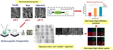
Hydroxyapatite nanoparticles (HAP NPs) with distinct morphologies were synthesized by the wet precipitation method by varying pH, and their shape was confirmed by scanning electron microscopy as spherical (pH 12), rod-like (pH 9), and needle-like (pH 8). The particle sizes of HAP NPs were 96.86 ± 1.48 nm for needle-shaped, 118 ± 4.32 nm for rod-shaped, and 94.43 ± 1.02 nm for spherical-shaped particles. XRD analysis showed clear and distinct peaks indicating crystalline nature, while FTIR confirmed the characteristic features of hydroxyapatite. The negative zeta potential of the HAP NPs was attributed to the presence of surface phosphate ions. The influence of HAP NP shape and size on intracellular uptake was evaluated in the MG-63 osteosarcoma cell line by Confocal Laser Scanning Microscopy (CLSM). CLSM results demonstrated that rod-shaped HAP NPs predominantly localized within the lysosome and nucleus, while spherical HAP NPs accumulated at the cell membrane. The MTT, clonogenic survival, cell scratch and transwell migration assays revealed that rod-shaped HAP NPs exhibited superior anticancer activity compared to their needle- and spherical-shaped counterparts and completely suppressed the clonogenic survival of MG-63 cells. Our findings confirm that the shape of HAP NPs is a critical factor influencing their intracellular uptake and anticancer activity.

 求助内容:
求助内容: 应助结果提醒方式:


