{"title":"松果体区原发性恶性黑色素瘤:说明性病例。","authors":"Jiahong Xu, Jibo Hu, Zhenwei Chen","doi":"10.3171/CASE25242","DOIUrl":null,"url":null,"abstract":"<p><strong>Background: </strong>Primary pineal melanoma is a rare tumor characterized by a high propensity for progression and recurrence. The imaging complexity of the melanoma will pose significant challenges for preoperative diagnosis. Definitive diagnosis necessitates histopathological examination.</p><p><strong>Observations: </strong>The authors present the case of a 49-year-old woman presenting with headache accompanied by nausea and vomiting. Imaging revealed a pineal tumor with subarachnoid hemorrhage. She underwent her first craniotomy at another hospital. Four months after surgery, she was sent to the authors' hospital for a second craniotomy due to recurrent cerebral hemorrhage. The postoperative pathological diagnosis was malignant melanoma in the pineal region.</p><p><strong>Lessons: </strong>In this case, malignant melanoma in the pineal region recurred rapidly and involved the ventricles only 4 months after surgery, suggesting the necessity of close follow-up for rare tumors. In addition, diagnosing this type of tumor on preoperative imaging is challenging, and although pathological diagnosis is the gold standard, the patient in this case required repeat pathological analysis for a definitive diagnosis to be made. https://thejns.org/doi/10.3171/CASE25242.</p>","PeriodicalId":94098,"journal":{"name":"Journal of neurosurgery. Case lessons","volume":"10 11","pages":""},"PeriodicalIF":0.0000,"publicationDate":"2025-09-15","publicationTypes":"Journal Article","fieldsOfStudy":null,"isOpenAccess":false,"openAccessPdf":"https://www.ncbi.nlm.nih.gov/pmc/articles/PMC12435373/pdf/","citationCount":"0","resultStr":"{\"title\":\"Primary malignant melanoma of the pineal region: illustrative case.\",\"authors\":\"Jiahong Xu, Jibo Hu, Zhenwei Chen\",\"doi\":\"10.3171/CASE25242\",\"DOIUrl\":null,\"url\":null,\"abstract\":\"<p><strong>Background: </strong>Primary pineal melanoma is a rare tumor characterized by a high propensity for progression and recurrence. The imaging complexity of the melanoma will pose significant challenges for preoperative diagnosis. Definitive diagnosis necessitates histopathological examination.</p><p><strong>Observations: </strong>The authors present the case of a 49-year-old woman presenting with headache accompanied by nausea and vomiting. Imaging revealed a pineal tumor with subarachnoid hemorrhage. She underwent her first craniotomy at another hospital. Four months after surgery, she was sent to the authors' hospital for a second craniotomy due to recurrent cerebral hemorrhage. The postoperative pathological diagnosis was malignant melanoma in the pineal region.</p><p><strong>Lessons: </strong>In this case, malignant melanoma in the pineal region recurred rapidly and involved the ventricles only 4 months after surgery, suggesting the necessity of close follow-up for rare tumors. In addition, diagnosing this type of tumor on preoperative imaging is challenging, and although pathological diagnosis is the gold standard, the patient in this case required repeat pathological analysis for a definitive diagnosis to be made. https://thejns.org/doi/10.3171/CASE25242.</p>\",\"PeriodicalId\":94098,\"journal\":{\"name\":\"Journal of neurosurgery. Case lessons\",\"volume\":\"10 11\",\"pages\":\"\"},\"PeriodicalIF\":0.0000,\"publicationDate\":\"2025-09-15\",\"publicationTypes\":\"Journal Article\",\"fieldsOfStudy\":null,\"isOpenAccess\":false,\"openAccessPdf\":\"https://www.ncbi.nlm.nih.gov/pmc/articles/PMC12435373/pdf/\",\"citationCount\":\"0\",\"resultStr\":null,\"platform\":\"Semanticscholar\",\"paperid\":null,\"PeriodicalName\":\"Journal of neurosurgery. Case lessons\",\"FirstCategoryId\":\"1085\",\"ListUrlMain\":\"https://doi.org/10.3171/CASE25242\",\"RegionNum\":0,\"RegionCategory\":null,\"ArticlePicture\":[],\"TitleCN\":null,\"AbstractTextCN\":null,\"PMCID\":null,\"EPubDate\":\"\",\"PubModel\":\"\",\"JCR\":\"\",\"JCRName\":\"\",\"Score\":null,\"Total\":0}","platform":"Semanticscholar","paperid":null,"PeriodicalName":"Journal of neurosurgery. Case lessons","FirstCategoryId":"1085","ListUrlMain":"https://doi.org/10.3171/CASE25242","RegionNum":0,"RegionCategory":null,"ArticlePicture":[],"TitleCN":null,"AbstractTextCN":null,"PMCID":null,"EPubDate":"","PubModel":"","JCR":"","JCRName":"","Score":null,"Total":0}
Primary malignant melanoma of the pineal region: illustrative case.
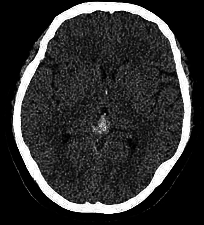
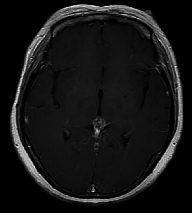
Background: Primary pineal melanoma is a rare tumor characterized by a high propensity for progression and recurrence. The imaging complexity of the melanoma will pose significant challenges for preoperative diagnosis. Definitive diagnosis necessitates histopathological examination.
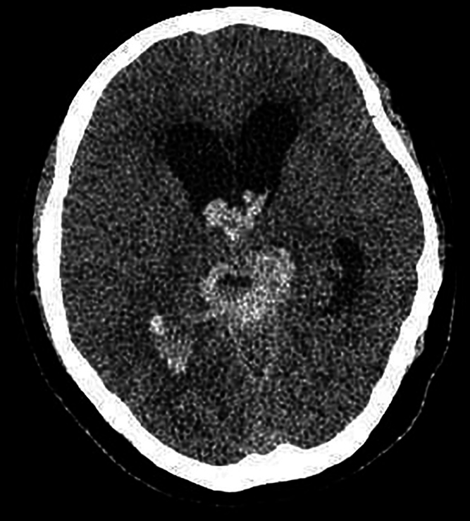
Observations: The authors present the case of a 49-year-old woman presenting with headache accompanied by nausea and vomiting. Imaging revealed a pineal tumor with subarachnoid hemorrhage. She underwent her first craniotomy at another hospital. Four months after surgery, she was sent to the authors' hospital for a second craniotomy due to recurrent cerebral hemorrhage. The postoperative pathological diagnosis was malignant melanoma in the pineal region.
Lessons: In this case, malignant melanoma in the pineal region recurred rapidly and involved the ventricles only 4 months after surgery, suggesting the necessity of close follow-up for rare tumors. In addition, diagnosing this type of tumor on preoperative imaging is challenging, and although pathological diagnosis is the gold standard, the patient in this case required repeat pathological analysis for a definitive diagnosis to be made. https://thejns.org/doi/10.3171/CASE25242.

 求助内容:
求助内容: 应助结果提醒方式:


