Derya Tireli, Jeppe Romme Christensen, Tina Nørgaard Munch, Nanna MacAulay, Henrik Bo Wiberg Larsson, Jonathan Frederik Carlsen, Stig Praestekjaer Cramer
{"title":"脉络膜丛容积在脑部疾病中的应用:系统综述。","authors":"Derya Tireli, Jeppe Romme Christensen, Tina Nørgaard Munch, Nanna MacAulay, Henrik Bo Wiberg Larsson, Jonathan Frederik Carlsen, Stig Praestekjaer Cramer","doi":"10.1186/s12987-025-00702-4","DOIUrl":null,"url":null,"abstract":"<p><strong>Background: </strong>The choroid plexus is a highly vascularized structure located in the lateral, third, and fourth ventricles of the brain. Recent studies suggest that volumetric changes in choroid plexus volume are associated with progression in various brain diseases. Segmentation algorithms have significantly improved our ability to study choroid plexus volumetrics in relation to various pathologies. Thus, the specific purpose of this review was to describe to what extent choroid plexus volume estimation provides clinically relevant information in brain diseases.</p><p><strong>Methods: </strong>An extensive literature search was conducted across Pubmed, Embase and Cochrane databases. A comprehensive, detailed qualitative descriptive analysis, and a thorough risk-of-bias assessment were performed for the included studies.</p><p><strong>Results: </strong>Forty-eight studies were included in this systematic review in the categories of multiple sclerosis, neurodegenerative diseases, psychiatric disorders, healthy populations and a group categorized as \"other\" for all other brain diseases that did not fit into the other categories.</p><p><strong>Conclusion: </strong>For many of the studies included, the patients had a larger choroid plexus volume compared to healthy controls. Evidence is currently insufficient to determine whether CPV enlargement correlates with clinical severity or functional scores. The most common segmentation technique was the automatic segmentation method, followed by manual correction of the segmented choroid plexus. Thus, this review highlights the growing interest choroid plexus volume, its segmentation, and its potential as a biomarker for numerous brain diseases.</p>","PeriodicalId":12321,"journal":{"name":"Fluids and Barriers of the CNS","volume":"22 1","pages":"92"},"PeriodicalIF":6.2000,"publicationDate":"2025-09-15","publicationTypes":"Journal Article","fieldsOfStudy":null,"isOpenAccess":false,"openAccessPdf":"https://www.ncbi.nlm.nih.gov/pmc/articles/PMC12439388/pdf/","citationCount":"0","resultStr":"{\"title\":\"Choroid plexus volume in brain disorders: a systematic review.\",\"authors\":\"Derya Tireli, Jeppe Romme Christensen, Tina Nørgaard Munch, Nanna MacAulay, Henrik Bo Wiberg Larsson, Jonathan Frederik Carlsen, Stig Praestekjaer Cramer\",\"doi\":\"10.1186/s12987-025-00702-4\",\"DOIUrl\":null,\"url\":null,\"abstract\":\"<p><strong>Background: </strong>The choroid plexus is a highly vascularized structure located in the lateral, third, and fourth ventricles of the brain. Recent studies suggest that volumetric changes in choroid plexus volume are associated with progression in various brain diseases. Segmentation algorithms have significantly improved our ability to study choroid plexus volumetrics in relation to various pathologies. Thus, the specific purpose of this review was to describe to what extent choroid plexus volume estimation provides clinically relevant information in brain diseases.</p><p><strong>Methods: </strong>An extensive literature search was conducted across Pubmed, Embase and Cochrane databases. A comprehensive, detailed qualitative descriptive analysis, and a thorough risk-of-bias assessment were performed for the included studies.</p><p><strong>Results: </strong>Forty-eight studies were included in this systematic review in the categories of multiple sclerosis, neurodegenerative diseases, psychiatric disorders, healthy populations and a group categorized as \\\"other\\\" for all other brain diseases that did not fit into the other categories.</p><p><strong>Conclusion: </strong>For many of the studies included, the patients had a larger choroid plexus volume compared to healthy controls. Evidence is currently insufficient to determine whether CPV enlargement correlates with clinical severity or functional scores. The most common segmentation technique was the automatic segmentation method, followed by manual correction of the segmented choroid plexus. Thus, this review highlights the growing interest choroid plexus volume, its segmentation, and its potential as a biomarker for numerous brain diseases.</p>\",\"PeriodicalId\":12321,\"journal\":{\"name\":\"Fluids and Barriers of the CNS\",\"volume\":\"22 1\",\"pages\":\"92\"},\"PeriodicalIF\":6.2000,\"publicationDate\":\"2025-09-15\",\"publicationTypes\":\"Journal Article\",\"fieldsOfStudy\":null,\"isOpenAccess\":false,\"openAccessPdf\":\"https://www.ncbi.nlm.nih.gov/pmc/articles/PMC12439388/pdf/\",\"citationCount\":\"0\",\"resultStr\":null,\"platform\":\"Semanticscholar\",\"paperid\":null,\"PeriodicalName\":\"Fluids and Barriers of the CNS\",\"FirstCategoryId\":\"3\",\"ListUrlMain\":\"https://doi.org/10.1186/s12987-025-00702-4\",\"RegionNum\":1,\"RegionCategory\":\"医学\",\"ArticlePicture\":[],\"TitleCN\":null,\"AbstractTextCN\":null,\"PMCID\":null,\"EPubDate\":\"\",\"PubModel\":\"\",\"JCR\":\"Q1\",\"JCRName\":\"NEUROSCIENCES\",\"Score\":null,\"Total\":0}","platform":"Semanticscholar","paperid":null,"PeriodicalName":"Fluids and Barriers of the CNS","FirstCategoryId":"3","ListUrlMain":"https://doi.org/10.1186/s12987-025-00702-4","RegionNum":1,"RegionCategory":"医学","ArticlePicture":[],"TitleCN":null,"AbstractTextCN":null,"PMCID":null,"EPubDate":"","PubModel":"","JCR":"Q1","JCRName":"NEUROSCIENCES","Score":null,"Total":0}
Choroid plexus volume in brain disorders: a systematic review.
Background: The choroid plexus is a highly vascularized structure located in the lateral, third, and fourth ventricles of the brain. Recent studies suggest that volumetric changes in choroid plexus volume are associated with progression in various brain diseases. Segmentation algorithms have significantly improved our ability to study choroid plexus volumetrics in relation to various pathologies. Thus, the specific purpose of this review was to describe to what extent choroid plexus volume estimation provides clinically relevant information in brain diseases.
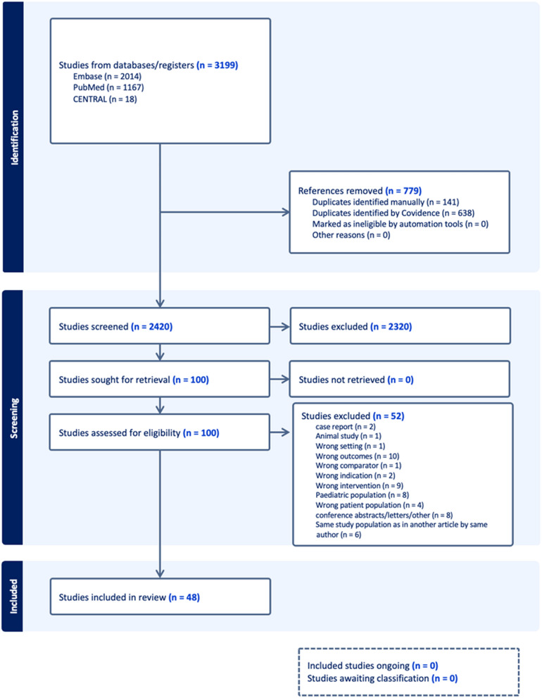
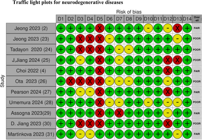
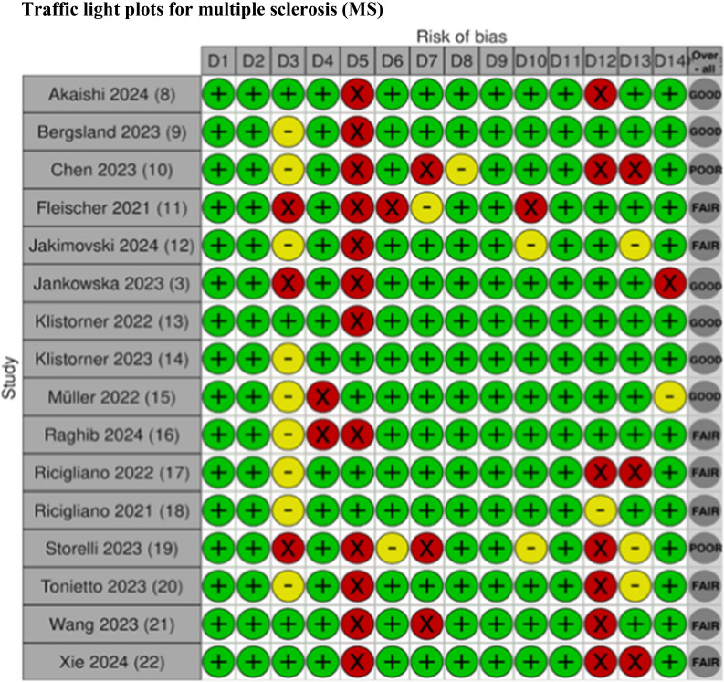
Methods: An extensive literature search was conducted across Pubmed, Embase and Cochrane databases. A comprehensive, detailed qualitative descriptive analysis, and a thorough risk-of-bias assessment were performed for the included studies.
Results: Forty-eight studies were included in this systematic review in the categories of multiple sclerosis, neurodegenerative diseases, psychiatric disorders, healthy populations and a group categorized as "other" for all other brain diseases that did not fit into the other categories.
Conclusion: For many of the studies included, the patients had a larger choroid plexus volume compared to healthy controls. Evidence is currently insufficient to determine whether CPV enlargement correlates with clinical severity or functional scores. The most common segmentation technique was the automatic segmentation method, followed by manual correction of the segmented choroid plexus. Thus, this review highlights the growing interest choroid plexus volume, its segmentation, and its potential as a biomarker for numerous brain diseases.
期刊介绍:
"Fluids and Barriers of the CNS" is a scholarly open access journal that specializes in the intricate world of the central nervous system's fluids and barriers, which are pivotal for the health and well-being of the human body. This journal is a peer-reviewed platform that welcomes research manuscripts exploring the full spectrum of CNS fluids and barriers, with a particular focus on their roles in both health and disease.
At the heart of this journal's interest is the cerebrospinal fluid (CSF), a vital fluid that circulates within the brain and spinal cord, playing a multifaceted role in the normal functioning of the brain and in various neurological conditions. The journal delves into the composition, circulation, and absorption of CSF, as well as its relationship with the parenchymal interstitial fluid and the neurovascular unit at the blood-brain barrier (BBB).
 求助内容:
求助内容: 应助结果提醒方式:


