Man-Ning Wu, Yue-Min Zou, Xiang-Nan Zhou, Sangwon Hong, Lei Wang, Yan-Ping Bai
{"title":"PFN1高表达的MAIT细胞介导银屑病的免疫激活和代谢重编程。","authors":"Man-Ning Wu, Yue-Min Zou, Xiang-Nan Zhou, Sangwon Hong, Lei Wang, Yan-Ping Bai","doi":"10.2147/CCID.S535795","DOIUrl":null,"url":null,"abstract":"<p><strong>Background: </strong>Psoriasis is a chronic inflammatory skin disease involving dysregulated immune responses and complex genetic factors. This study combines single-cell RNA sequencing (scRNA-seq), gene expression profiling, and genetic analysis to explore cellular and molecular contributors to psoriasis.</p><p><strong>Methods: </strong>Single-cell RNA-seq data (n = 3 psoriasis, n = 2 control; GSE228421) were used for cell-type annotation and functional characterization. T cell subsets were analyzed for differentiation trajectories and cell-cell communication. Differentially expressed genes in mucosal-associated invariant T (MAIT) cells were evaluated by enrichment analysis. Candidate gene causality was tested via eQTL-based Mendelian randomization (MR) and supported by bulk RNA-seq validation.</p><p><strong>Results: </strong>MAIT cells were enriched in psoriatic lesions and exhibited strong intercellular interactions. Functional analyses revealed activation of IL6-JAK-STAT3 signaling, TNF-NFκB pathway, and glycolysis in MAIT cells. MR identified RPS20 as a protective factor (OR = 0.5994, p = 0.011) and PFN1 as a potential risk gene (OR = 1.7229, p = 0.037), with PFN1 highly expressed in MAIT cells. Colocalization analysis showed no significant genetic overlap between PFN1 expression and psoriasis risk. Metabolic profiling revealed differential pathway involvement in PFN1+ and PFN1- MAIT cells.</p><p><strong>Conclusion: </strong>Our integrative analysis highlights MAIT cells and PFN1 as likely contributors to psoriasis pathogenesis. These findings offer insights into immune and metabolic alterations, suggesting potential targets for therapeutic intervention.</p>","PeriodicalId":10447,"journal":{"name":"Clinical, Cosmetic and Investigational Dermatology","volume":"18 ","pages":"2243-2257"},"PeriodicalIF":2.2000,"publicationDate":"2025-09-09","publicationTypes":"Journal Article","fieldsOfStudy":null,"isOpenAccess":false,"openAccessPdf":"https://www.ncbi.nlm.nih.gov/pmc/articles/PMC12433214/pdf/","citationCount":"0","resultStr":"{\"title\":\"MAIT Cells with High PFN1 Expression Mediate Immune Activation and Metabolic Reprogramming in Psoriasis.\",\"authors\":\"Man-Ning Wu, Yue-Min Zou, Xiang-Nan Zhou, Sangwon Hong, Lei Wang, Yan-Ping Bai\",\"doi\":\"10.2147/CCID.S535795\",\"DOIUrl\":null,\"url\":null,\"abstract\":\"<p><strong>Background: </strong>Psoriasis is a chronic inflammatory skin disease involving dysregulated immune responses and complex genetic factors. This study combines single-cell RNA sequencing (scRNA-seq), gene expression profiling, and genetic analysis to explore cellular and molecular contributors to psoriasis.</p><p><strong>Methods: </strong>Single-cell RNA-seq data (n = 3 psoriasis, n = 2 control; GSE228421) were used for cell-type annotation and functional characterization. T cell subsets were analyzed for differentiation trajectories and cell-cell communication. Differentially expressed genes in mucosal-associated invariant T (MAIT) cells were evaluated by enrichment analysis. Candidate gene causality was tested via eQTL-based Mendelian randomization (MR) and supported by bulk RNA-seq validation.</p><p><strong>Results: </strong>MAIT cells were enriched in psoriatic lesions and exhibited strong intercellular interactions. Functional analyses revealed activation of IL6-JAK-STAT3 signaling, TNF-NFκB pathway, and glycolysis in MAIT cells. MR identified RPS20 as a protective factor (OR = 0.5994, p = 0.011) and PFN1 as a potential risk gene (OR = 1.7229, p = 0.037), with PFN1 highly expressed in MAIT cells. Colocalization analysis showed no significant genetic overlap between PFN1 expression and psoriasis risk. Metabolic profiling revealed differential pathway involvement in PFN1+ and PFN1- MAIT cells.</p><p><strong>Conclusion: </strong>Our integrative analysis highlights MAIT cells and PFN1 as likely contributors to psoriasis pathogenesis. These findings offer insights into immune and metabolic alterations, suggesting potential targets for therapeutic intervention.</p>\",\"PeriodicalId\":10447,\"journal\":{\"name\":\"Clinical, Cosmetic and Investigational Dermatology\",\"volume\":\"18 \",\"pages\":\"2243-2257\"},\"PeriodicalIF\":2.2000,\"publicationDate\":\"2025-09-09\",\"publicationTypes\":\"Journal Article\",\"fieldsOfStudy\":null,\"isOpenAccess\":false,\"openAccessPdf\":\"https://www.ncbi.nlm.nih.gov/pmc/articles/PMC12433214/pdf/\",\"citationCount\":\"0\",\"resultStr\":null,\"platform\":\"Semanticscholar\",\"paperid\":null,\"PeriodicalName\":\"Clinical, Cosmetic and Investigational Dermatology\",\"FirstCategoryId\":\"3\",\"ListUrlMain\":\"https://doi.org/10.2147/CCID.S535795\",\"RegionNum\":4,\"RegionCategory\":\"医学\",\"ArticlePicture\":[],\"TitleCN\":null,\"AbstractTextCN\":null,\"PMCID\":null,\"EPubDate\":\"2025/1/1 0:00:00\",\"PubModel\":\"eCollection\",\"JCR\":\"Q3\",\"JCRName\":\"DERMATOLOGY\",\"Score\":null,\"Total\":0}","platform":"Semanticscholar","paperid":null,"PeriodicalName":"Clinical, Cosmetic and Investigational Dermatology","FirstCategoryId":"3","ListUrlMain":"https://doi.org/10.2147/CCID.S535795","RegionNum":4,"RegionCategory":"医学","ArticlePicture":[],"TitleCN":null,"AbstractTextCN":null,"PMCID":null,"EPubDate":"2025/1/1 0:00:00","PubModel":"eCollection","JCR":"Q3","JCRName":"DERMATOLOGY","Score":null,"Total":0}
引用次数: 0
摘要
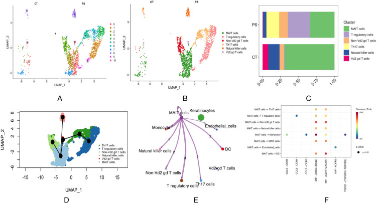
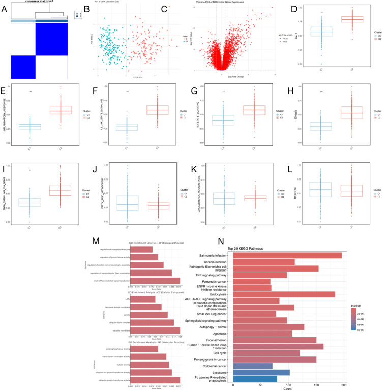
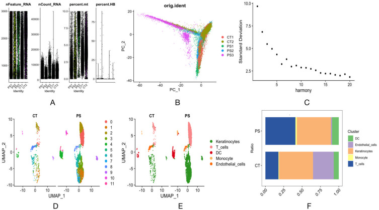
背景:银屑病是一种慢性炎症性皮肤病,涉及免疫反应失调和复杂的遗传因素。本研究结合单细胞RNA测序(scRNA-seq)、基因表达谱和遗传分析来探索银屑病的细胞和分子因素。方法:使用单细胞RNA-seq数据(n = 3例牛皮癣患者,n = 2例对照组;GSE228421)进行细胞类型注释和功能表征。分析T细胞亚群的分化轨迹和细胞间通讯。通过富集分析评估粘膜相关不变T (MAIT)细胞中的差异表达基因。通过基于eqtl的孟德尔随机化(MR)测试候选基因的因果关系,并通过大量RNA-seq验证进行支持。结果:MAIT细胞在银屑病病变中富集,并表现出强烈的细胞间相互作用。功能分析显示,在MAIT细胞中,IL6-JAK-STAT3信号通路、TNF-NFκB通路和糖酵解被激活。MR鉴定RPS20为保护因子(OR = 0.5994, p = 0.011), PFN1为潜在危险基因(OR = 1.7229, p = 0.037), PFN1在MAIT细胞中高表达。共定位分析显示PFN1表达与牛皮癣风险之间没有明显的遗传重叠。代谢谱显示PFN1+和PFN1- MAIT细胞参与了不同的通路。结论:我们的综合分析强调MAIT细胞和PFN1可能是银屑病发病机制的贡献者。这些发现提供了对免疫和代谢改变的见解,提出了治疗干预的潜在目标。
MAIT Cells with High PFN1 Expression Mediate Immune Activation and Metabolic Reprogramming in Psoriasis.
Background: Psoriasis is a chronic inflammatory skin disease involving dysregulated immune responses and complex genetic factors. This study combines single-cell RNA sequencing (scRNA-seq), gene expression profiling, and genetic analysis to explore cellular and molecular contributors to psoriasis.
Methods: Single-cell RNA-seq data (n = 3 psoriasis, n = 2 control; GSE228421) were used for cell-type annotation and functional characterization. T cell subsets were analyzed for differentiation trajectories and cell-cell communication. Differentially expressed genes in mucosal-associated invariant T (MAIT) cells were evaluated by enrichment analysis. Candidate gene causality was tested via eQTL-based Mendelian randomization (MR) and supported by bulk RNA-seq validation.
Results: MAIT cells were enriched in psoriatic lesions and exhibited strong intercellular interactions. Functional analyses revealed activation of IL6-JAK-STAT3 signaling, TNF-NFκB pathway, and glycolysis in MAIT cells. MR identified RPS20 as a protective factor (OR = 0.5994, p = 0.011) and PFN1 as a potential risk gene (OR = 1.7229, p = 0.037), with PFN1 highly expressed in MAIT cells. Colocalization analysis showed no significant genetic overlap between PFN1 expression and psoriasis risk. Metabolic profiling revealed differential pathway involvement in PFN1+ and PFN1- MAIT cells.
Conclusion: Our integrative analysis highlights MAIT cells and PFN1 as likely contributors to psoriasis pathogenesis. These findings offer insights into immune and metabolic alterations, suggesting potential targets for therapeutic intervention.
期刊介绍:
Clinical, Cosmetic and Investigational Dermatology is an international, peer-reviewed, open access journal that focuses on the latest clinical and experimental research in all aspects of skin disease and cosmetic interventions. Normal and pathological processes in skin development and aging, their modification and treatment, as well as basic research into histology of dermal and dermal structures that provide clinical insights and potential treatment options are key topics for the journal.
Patient satisfaction, preference, quality of life, compliance, persistence and their role in developing new management options to optimize outcomes for target conditions constitute major areas of interest.
The journal is characterized by the rapid reporting of clinical studies, reviews and original research in skin research and skin care.
All areas of dermatology will be covered; contributions will be welcomed from all clinicians and basic science researchers globally.
 求助内容:
求助内容: 应助结果提醒方式:


