LaToya McLean, Carrie Andrews, Louis Cappelli, Grant Gillan, Mark Curtis, James J Evans, Wenyin Shi
{"title":"垂体大腺瘤和硬瘤性颅咽管瘤:鞍部碰撞肿瘤1例报告。","authors":"LaToya McLean, Carrie Andrews, Louis Cappelli, Grant Gillan, Mark Curtis, James J Evans, Wenyin Shi","doi":"10.1155/crnm/6895334","DOIUrl":null,"url":null,"abstract":"<p><p>We present a rare case of a collision tumor involving a pituitary macroadenoma and adamantinomatous craniopharyngioma in a 49-year-old woman. The patient presented with a 2-year history of amenorrhea and elevated prolactin. Brain MRI revealed two sellar masses. Initially managed with observation due to the absence of neurological deficits, surgical resection was later performed after clinical and radiographic progression. Pathology confirmed both tumor types: pituitary macroadenoma and adamantinomatous craniopharyngioma. Postoperative MRI showed residual disease at the superior margin. The patient subsequently received fractionated stereotactic radiation for residual disease and tolerated well.</p>","PeriodicalId":9615,"journal":{"name":"Case Reports in Neurological Medicine","volume":"2025 ","pages":"6895334"},"PeriodicalIF":0.9000,"publicationDate":"2025-09-04","publicationTypes":"Journal Article","fieldsOfStudy":null,"isOpenAccess":false,"openAccessPdf":"https://www.ncbi.nlm.nih.gov/pmc/articles/PMC12425626/pdf/","citationCount":"0","resultStr":"{\"title\":\"Pituitary Macroadenoma and Adamantinomatous Craniopharyngioma: A Rare Case Report of Sellar Collision Tumors.\",\"authors\":\"LaToya McLean, Carrie Andrews, Louis Cappelli, Grant Gillan, Mark Curtis, James J Evans, Wenyin Shi\",\"doi\":\"10.1155/crnm/6895334\",\"DOIUrl\":null,\"url\":null,\"abstract\":\"<p><p>We present a rare case of a collision tumor involving a pituitary macroadenoma and adamantinomatous craniopharyngioma in a 49-year-old woman. The patient presented with a 2-year history of amenorrhea and elevated prolactin. Brain MRI revealed two sellar masses. Initially managed with observation due to the absence of neurological deficits, surgical resection was later performed after clinical and radiographic progression. Pathology confirmed both tumor types: pituitary macroadenoma and adamantinomatous craniopharyngioma. Postoperative MRI showed residual disease at the superior margin. The patient subsequently received fractionated stereotactic radiation for residual disease and tolerated well.</p>\",\"PeriodicalId\":9615,\"journal\":{\"name\":\"Case Reports in Neurological Medicine\",\"volume\":\"2025 \",\"pages\":\"6895334\"},\"PeriodicalIF\":0.9000,\"publicationDate\":\"2025-09-04\",\"publicationTypes\":\"Journal Article\",\"fieldsOfStudy\":null,\"isOpenAccess\":false,\"openAccessPdf\":\"https://www.ncbi.nlm.nih.gov/pmc/articles/PMC12425626/pdf/\",\"citationCount\":\"0\",\"resultStr\":null,\"platform\":\"Semanticscholar\",\"paperid\":null,\"PeriodicalName\":\"Case Reports in Neurological Medicine\",\"FirstCategoryId\":\"1085\",\"ListUrlMain\":\"https://doi.org/10.1155/crnm/6895334\",\"RegionNum\":0,\"RegionCategory\":null,\"ArticlePicture\":[],\"TitleCN\":null,\"AbstractTextCN\":null,\"PMCID\":null,\"EPubDate\":\"2025/1/1 0:00:00\",\"PubModel\":\"eCollection\",\"JCR\":\"Q4\",\"JCRName\":\"CLINICAL NEUROLOGY\",\"Score\":null,\"Total\":0}","platform":"Semanticscholar","paperid":null,"PeriodicalName":"Case Reports in Neurological Medicine","FirstCategoryId":"1085","ListUrlMain":"https://doi.org/10.1155/crnm/6895334","RegionNum":0,"RegionCategory":null,"ArticlePicture":[],"TitleCN":null,"AbstractTextCN":null,"PMCID":null,"EPubDate":"2025/1/1 0:00:00","PubModel":"eCollection","JCR":"Q4","JCRName":"CLINICAL NEUROLOGY","Score":null,"Total":0}
Pituitary Macroadenoma and Adamantinomatous Craniopharyngioma: A Rare Case Report of Sellar Collision Tumors.
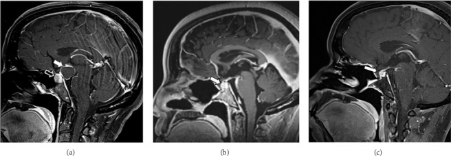
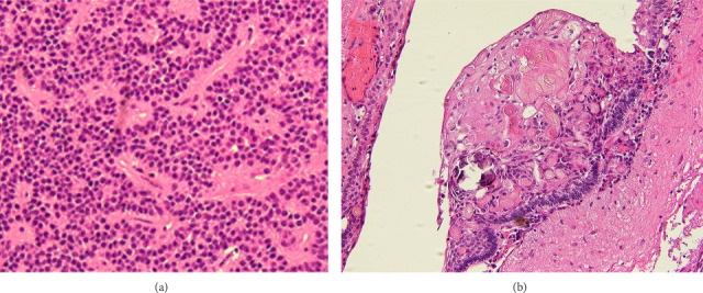
We present a rare case of a collision tumor involving a pituitary macroadenoma and adamantinomatous craniopharyngioma in a 49-year-old woman. The patient presented with a 2-year history of amenorrhea and elevated prolactin. Brain MRI revealed two sellar masses. Initially managed with observation due to the absence of neurological deficits, surgical resection was later performed after clinical and radiographic progression. Pathology confirmed both tumor types: pituitary macroadenoma and adamantinomatous craniopharyngioma. Postoperative MRI showed residual disease at the superior margin. The patient subsequently received fractionated stereotactic radiation for residual disease and tolerated well.

 求助内容:
求助内容: 应助结果提醒方式:


