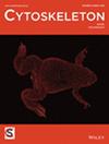
{"title":"封底内图","authors":"","doi":"10.1002/cm.70038","DOIUrl":null,"url":null,"abstract":"<p>ON THE INNER BACK COVER: Maximum projection image of mouse tenocytes treated with TGF2. Nuclei are labeled with Hoechst (magenta) and filamentous actin (F-actin) is stained with Rhodamine—Phalloidin (cyan to white).</p><p>Credit: Rylee E. King (University of Delaware, USA)\n\n <figure>\n <div><picture>\n <source></source></picture><p></p>\n </div>\n </figure>\n </p>","PeriodicalId":55186,"journal":{"name":"Cytoskeleton","volume":"82 9","pages":""},"PeriodicalIF":1.6000,"publicationDate":"2025-09-11","publicationTypes":"Journal Article","fieldsOfStudy":null,"isOpenAccess":false,"openAccessPdf":"https://onlinelibrary.wiley.com/doi/epdf/10.1002/cm.70038","citationCount":"0","resultStr":"{\"title\":\"Inner Back Cover Image\",\"authors\":\"\",\"doi\":\"10.1002/cm.70038\",\"DOIUrl\":null,\"url\":null,\"abstract\":\"<p>ON THE INNER BACK COVER: Maximum projection image of mouse tenocytes treated with TGF2. Nuclei are labeled with Hoechst (magenta) and filamentous actin (F-actin) is stained with Rhodamine—Phalloidin (cyan to white).</p><p>Credit: Rylee E. King (University of Delaware, USA)\\n\\n <figure>\\n <div><picture>\\n <source></source></picture><p></p>\\n </div>\\n </figure>\\n </p>\",\"PeriodicalId\":55186,\"journal\":{\"name\":\"Cytoskeleton\",\"volume\":\"82 9\",\"pages\":\"\"},\"PeriodicalIF\":1.6000,\"publicationDate\":\"2025-09-11\",\"publicationTypes\":\"Journal Article\",\"fieldsOfStudy\":null,\"isOpenAccess\":false,\"openAccessPdf\":\"https://onlinelibrary.wiley.com/doi/epdf/10.1002/cm.70038\",\"citationCount\":\"0\",\"resultStr\":null,\"platform\":\"Semanticscholar\",\"paperid\":null,\"PeriodicalName\":\"Cytoskeleton\",\"FirstCategoryId\":\"99\",\"ListUrlMain\":\"https://onlinelibrary.wiley.com/doi/10.1002/cm.70038\",\"RegionNum\":4,\"RegionCategory\":\"生物学\",\"ArticlePicture\":[],\"TitleCN\":null,\"AbstractTextCN\":null,\"PMCID\":null,\"EPubDate\":\"\",\"PubModel\":\"\",\"JCR\":\"Q4\",\"JCRName\":\"CELL BIOLOGY\",\"Score\":null,\"Total\":0}","platform":"Semanticscholar","paperid":null,"PeriodicalName":"Cytoskeleton","FirstCategoryId":"99","ListUrlMain":"https://onlinelibrary.wiley.com/doi/10.1002/cm.70038","RegionNum":4,"RegionCategory":"生物学","ArticlePicture":[],"TitleCN":null,"AbstractTextCN":null,"PMCID":null,"EPubDate":"","PubModel":"","JCR":"Q4","JCRName":"CELL BIOLOGY","Score":null,"Total":0}
引用次数: 0
摘要
内后盖:TGF2处理后小鼠细胞的最大投影图像。细胞核用赫斯特(洋红色)标记,丝状肌动蛋白(f -肌动蛋白)用罗丹明- phalloidin(青色至白色)染色。来源:Rylee E. King(美国特拉华大学)
ON THE INNER BACK COVER: Maximum projection image of mouse tenocytes treated with TGF2. Nuclei are labeled with Hoechst (magenta) and filamentous actin (F-actin) is stained with Rhodamine—Phalloidin (cyan to white).
Credit: Rylee E. King (University of Delaware, USA)
期刊介绍:
Cytoskeleton focuses on all aspects of cytoskeletal research in healthy and diseased states, spanning genetic and cell biological observations, biochemical, biophysical and structural studies, mathematical modeling and theory. This includes, but is certainly not limited to, classic polymer systems of eukaryotic cells and their structural sites of attachment on membranes and organelles, as well as the bacterial cytoskeleton, the nucleoskeleton, and uncoventional polymer systems with structural/organizational roles. Cytoskeleton is published in 12 issues annually, and special issues will be dedicated to especially-active or newly-emerging areas of cytoskeletal research.